The Cerebellum: Brain for an Implicit Self (62 page)
Read The Cerebellum: Brain for an Implicit Self Online
Authors: Masao Ito
Tags: #Science, #Life Sciences, #Medical, #Biology, #Neurology, #Neuroscience

Szentágothai
, Janos,
28
task dependencies, functional stretch reflex
,
126
temporoparietal cortex
,
193
tendon tap
,
122
thought processes
activity in the cerebellum,
198-201
cerebellar internal model for thoughts,
196
cerebral cortical model for thoughts,
195-196
explicit/implicit thoughts,
197-198
mental disorders associated with cerebellar dysfunction,
201-203
neural systems,
193-195
throwing a ball, voluntary motor control
,
153-154
tools, motor actions
,
190-192
traditional views of cerebellum
involvement in motor skills,
25-27
morphological map,
22-25
Purkinje cells,
27-28
transcriptional inhibitors, protein synthesis
,
88
transduction pathways (conjunctive LTD)
,
71
AMPA receptors,
74-75
brain-derived neurotrophic factor (BDNF),
80
c-Fos/Jun-B,
79
Ca
2+
surge in Purkinje cell dendrites,
71-74
Ca
2+
/calmodulin-dependent protein kinase (CaMKII),
78
glial fibrillary acidic protein (GFAP),
79
glutamate receptor d2 (GluR d2),
76-77
insulin-like growth factor-1 (IGF-1),
80
Nitric oxide (NO) synthase,
75-76
PKCα and lipid-signaling cascade,
74
protein phosphatases,
78
protein tyrosine kinases,
77
translational inhibitors, protein synthesis
,
88
Tsukahara
, Nakakira,
30
unipolar brush cells
,
46-47
V-type microcomplex, adaptive control prototype
,
146-149
velocity storage, VOR (Vestibuloocular Reflex)
,
107
ventral spinocerebellar tract (VSCT)
,
60
vergence, frontal eye field
,
164-165
vermal lobules VI/VII, eye movements
,
159
vermis
,
23
vestibular compensation, VOR (Vestibuloocular Reflex)
,
112-113
vestibular mossy fiber input, VOR adaptation
,
110
vestibular nuclear neurons
,
68
vestibular organs
,
23
vestibulocerebellum
,
23
Vestibuloocular Reflex (VOR)
,
36
,
105-106
adaptation control system
climbing fiber input
,
110-111
eye movement-related signals
,
111-112
flocculus
,
107-109
memory sites
,
112
vestibular mossy fiber input
,
110
adaptive control,
139-140
feedforward control system,
39
neural integrator,
107
velocity storage,
107
vestibular compensation,
112-113
vestibulosympathetic reflex
,
136-137
voltage-gated Ca
2+
channels
,
71
voluntary eye movement
,
159
DMFC (dorsomedial frontal cortex),
165-166
frontal eye field,
159-165
voluntary motor control
,
150
hand grip,
154-156
instruction signals,
157-158
internal models,
167
internal forward model
,
167-175
internal inverse model
,
170-175
MOSAIC (modular selection and identification for control)
,
175
sensory cancellation
,
178-180
transition from reflex control to voluntary
,
176-179
load compensation,
150
multijoint arm movements,
151-154
operant conditioning,
156-157
reaction time,
151
voluntary movements
,
12
hybrid control,
13-14
motor actions,
18-19
VOR (Vestibuloocular Reflex)
,
36
,
105-106
adaptation control system
climbing fiber input
,
110-111
eye movement-related signals
,
111-112
flocculus
,
107-109
memory sites
,
112
vestibular mossy fiber input
,
110
adaptive control,
139-140
feedforward control system,
39
neural integrator,
107
velocity storage,
107
vestibular compensation,
112-113
VSCT (ventral spinocerebellar tract)
,
60
whale cerebellum
,
24-25
wiring diagrams
,
31
Wisconsin card-sorting test
,
199
XXVII Congress of the International Physiological Union
,
36
Yoshida
, Mitsuo,
30
zebrin
,
24
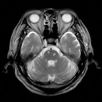
Color Plate I. MRI image of the human cerebellum.
Recent remarkable progress in brain imaging has made it possible to observe the human cerebellum in vivo. This horizontal section image was taken by the author on June 7, 2010. Abbreviations: b, basilar artery; h, cerebellar hemisphere; p, pons; v, 4th ventricle. For a three-dimensional atlas of the human cerebellum, see
Schmahmann et al., 1999
.

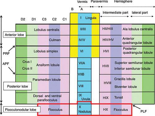
Color Plate II. A schematized map of the divisions of the cerebellar cortex.
On the outline of the anatomical divisions of the cerebellum (vermis, paravermis, and hemisphere) a lattice is formed by crossing transverse lobules and longitudinal zones to set coordinates for defining the position of each cortical area. Note that subzones A2, X, and Y are not shown. Note also that the zones and lobules are shown with the same width for simplicity despite that the fact that their widths vary considerably. The nomenclature used is that for the generalized mammalian cerebellum. Abbreviations: I-X, lobular divisions of the cerebellar vermis and paravermis; A-D2, longitudinal zones of the cerebellum; HI-HX, lobuli extending laterally from I-X. APF, ansoparamedian fissure; PFR, primary fissure; PLF, posterolateral fissure.

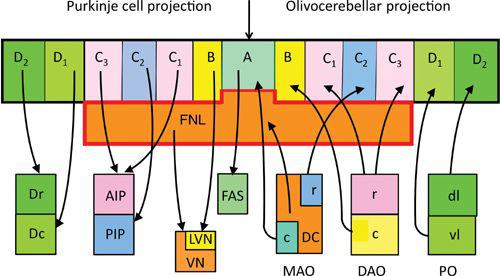
Color Plate III. Longitudinal zonal organization in the Purkinje cell output and climbing fiber input.
Upper half shows longitudinal zones and flocculonodular lobe after Color Plate II. Lower half, Target nuclei for Purkinje cell zones in the left half of the diagram. The right side shows the input connection from the inferior olive. Abbreviations: A-D
2
, zonal bands; AIP, anterior interpositus nucleus; c, caudal (MAO or DAO); DC, dorsal cap; Dc, caudal subnucleus of the dentate nucleus; Dr, rostral subnucleus of the dentate nucleus; dl, dorsal lamella; FAS, fastigial nucleus; FNL, flocculonodular lobe; LVN, lateral vestibular nucleus; MAO, medial accessory olive; PIP, posterior interpositus nucleus; PO, principal olive; r, rostral (MAO or DAO); VN, vestibular nuclei; vl, ventral lamella. (For further details, see
Voogd, 2010
.)

