Ross & Wilson Anatomy and Physiology in Health and Illness (160 page)
Read Ross & Wilson Anatomy and Physiology in Health and Illness Online
Authors: Anne Waugh,Allison Grant
Tags: #Medical, #Nursing, #General, #Anatomy

pH balance
In order to maintain the normal blood pH (acid–base balance), the cells of the proximal convoluted tubules secrete hydrogen ions. In the filtrate they combine with buffers (
p. 22
):
•
bicarbonate, forming carbonic acid

•
ammonia, forming ammonium ions

•
hydrogen phosphate, forming dihydrogen phosphate

Carbonic acid is converted to carbon dioxide (CO
2
) and water (H
2
O), and the CO
2
is reabsorbed, maintaining the buffering capacity of the blood. Hydrogen ions are excreted in the urine as ammonium salts and hydrogen phosphate. The normal pH of urine varies from 4.5 to 8 depending on diet, time of day and a number of other factors. Individuals whose diet contains a large amount of animal proteins tend to produce more acidic urine (lower pH) than vegetarians.
Ureters
Learning outcome
After studying this section you should be able to:
•
outline the structure and function of the ureters.
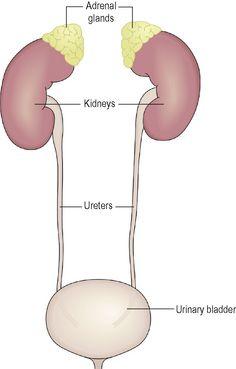
The ureters are the tubes that carry urine from the kidneys to the urinary bladder (
Fig. 13.17
). They are about 25 to 30 cm long with a diameter of about 3 mm.
Figure 13.17
The ureters and their relationship to the kidneys and bladder.
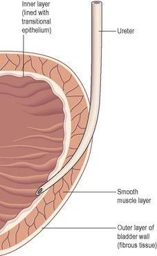
The ureter is continuous with the funnel-shaped renal pelvis. It passes downwards through the abdominal cavity, behind the peritoneum in front of the psoas muscle into the pelvic cavity, and passes obliquely through the posterior wall of the bladder (
Fig. 13.18
). Because of this arrangement, when urine accumulates and the pressure in the bladder rises, the ureters are compressed and the openings occluded. This prevents reflux of urine into the ureters (towards the kidneys) as the bladder fills and during micturition, when pressure increases as the muscular bladder wall contracts.
Figure 13.18
The position of the ureter where it passes through the bladder wall.
Structure
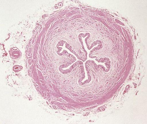
The walls of the ureters consist of three layers of tissue which are shown in cross-section in
Figure 13.19
:
•
an outer covering of
fibrous tissue
, continuous with the fibrous capsule of the kidney
•
a middle
muscular layer
consisting of interlacing smooth muscle fibres that form a functional unit round the ureter and an additional outer longitudinal layer in the lower third
•
an inner layer, the mucosa, composed of transitional epithelium (see
p. 35
).
Figure 13.19
Cross-section of the ureter.
Function
The ureters propel urine from the kidneys into the bladder by peristaltic contraction of the smooth muscle layer. This is an intrinsic property of the smooth muscle and is not under autonomic nerve control. Peristalsis originates in a pacemaker in the minor calyces. Peristaltic waves occur several times per minute, increasing in frequency with the volume of urine produced, sending little spurts of urine into the bladder.
Urinary bladder


