Ross & Wilson Anatomy and Physiology in Health and Illness (156 page)
Read Ross & Wilson Anatomy and Physiology in Health and Illness Online
Authors: Anne Waugh,Allison Grant
Tags: #Medical, #Nursing, #General, #Anatomy

Figure 13.1
The parts of the urinary system (excluding the urethra) and some associated structures.
The urinary system plays a vital part in maintaining homeostasis of water and electrolyte concentrations within the body. The kidneys produce urine that contains metabolic waste products, including the nitrogenous compounds urea and uric acid, excess ions and some drugs.
The main functions of the kidneys are:
•
formation and secretion of urine, which regulates total body water, electrolyte and acid–base balance and enables excretion of waste products
•
production and secretion of
erythropoietin
, the hormone that stimulates formation of red blood cells (
p. 60
)
•
production and secretion of
renin
, an important enzyme in the control of blood pressure (
p. 28
).
Urine is stored in the bladder and excreted by the process of
micturition
.
The first sections of this chapter explore the structures and functions of the organs described above. In the final section the consequences of abnormal functioning of the various parts of the urinary system on body function are considered.
Kidneys
Learning outcomes
After studying this section you should be able to:
identify the organs associated with the kidneys
outline the gross structure of the kidneys
describe the structure of a nephron
explain the processes involved in the formation of urine
explain how body water and electrolyte balance is maintained.
The kidneys (
Fig. 13.2
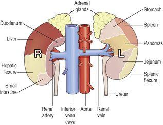
) lie on the posterior abdominal wall, one on each side of the vertebral column, behind the peritoneum and below the diaphragm. They extend from the level of the 12th thoracic vertebra to the 3rd lumbar vertebra, receiving some protection from the lower rib cage. The right kidney is usually slightly lower than the left, probably because of the considerable space occupied by the liver.
Figure 13.2
Anterior view of the kidneys showing the areas of contact with associated structures.
Kidneys are bean-shaped organs, about 11 cm long, 6 cm wide, 3 cm thick and weigh 150 g. They are embedded in, and held in position by, a mass of fat. A sheath of fibrous connective tissue, also known as the
renal fascia,
encloses the kidney and the renal fat.
Organs associated with the kidneys (
Figs 13.1
,
13.2
and
13.3
As the kidneys lie on either side of the vertebral column, each is associated with a different group of structures.
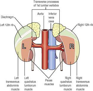
Figure 13.3
Posterior view of the kidneys showing the areas of contact with associated structures.
Right kidney
Superiorly
– the right adrenal gland
Anteriorly
– the right lobe of the liver, the duodenum and the hepatic flexure of the colon
Posteriorly
– the diaphragm, and muscles of the posterior abdominal wall
Left kidney
Superiorly
– the left adrenal gland
Anteriorly
– the spleen, stomach, pancreas, jejunum and splenic flexure of the colon
Posteriorly
– the diaphragm and muscles of the posterior abdominal wall
Gross structure of the kidney
There are three areas of tissue that can be distinguished when a longitudinal section of the kidney is viewed with the naked eye (
Fig. 13.4
):
•
an outer fibrous
capsule
, surrounding the kidney
•
the
cortex
, a reddish-brown layer of tissue immediately below the capsule and outside the pyramids
•
the
medulla
, the innermost layer, consisting of pale conical-shaped striations, the
renal pyramids
.


