Ross & Wilson Anatomy and Physiology in Health and Illness (111 page)
Read Ross & Wilson Anatomy and Physiology in Health and Illness Online
Authors: Anne Waugh,Allison Grant
Tags: #Medical, #Nursing, #General, #Anatomy

Figure 10.10
The trachea and some of its related structures.
Structures associated with the trachea (
Fig. 10.10
)
Superiorly
– the larynx
Inferiorly
– the right and left bronchi
Anteriorly
– upper part: the isthmus of the thyroid gland; lower part: the arch of the aorta and the sternum
Posteriorly
– the oesophagus separates the trachea from the vertebral column
Laterally
– the lungs and the lobes of the thyroid gland.
Structure
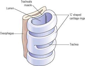
The trachea is composed of three layers of tissue, and held open by between 16 and 20 incomplete (C-shaped) rings of hyaline cartilage lying one above the other. The rings are incomplete posteriorly. The cartilages are embedded in a sleeve of smooth muscle and connective tissue, which also forms the posterior wall where the rings are incomplete. The soft tissue posterior wall is in contact with the oesophagus (
Fig. 10.11
).
Figure 10.11
The relationship of the trachea to the oesophagus.
Three layers of tissue ‘clothe’ the cartilages of the trachea.
•
The outer layer contains the fibrous and elastic tissue and encloses the cartilages.
•
The middle layer consists of cartilages and bands of smooth muscle that wind round the trachea in a helical arrangement. There is some areolar tissue, containing blood and lymph vessels and autonomic nerves. The free ends of the incomplete cartilages are connected by the trachealis muscle, which allows for adjustment of tracheal diameter.
•
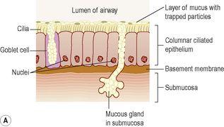
The lining is ciliated columnar epithelium, containing mucus-secreting goblet cells (
Fig. 10.12
).
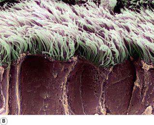
Figure 10.12
A.
Ciliated mucous membrane.
B.
Coloured scanning electron micrograph of bronchial cilia.
Blood and nerve supply, lymph drainage
The arterial blood supply is mainly by the inferior thyroid and bronchial arteries and the venous return is by the inferior thyroid veins into the brachiocephalic veins.
Parasympathetic nerve supply is by the recurrent laryngeal nerves and other branches of the vagi. Sympathetic supply is by nerves from the sympathetic ganglia. Parasympathetic stimulation constricts the trachea, and sympathetic stimulation dilates it.
Lymph from the respiratory passages drains through lymph nodes situated round the trachea and in the carina, the area where it divides into two bronchi.
Functions
Support and patency
Tracheal cartilages hold the trachea permanently open (patent), but the soft tissue bands in between the cartilages allow flexibility so that the head and neck can move freely without obstructing or kinking the trachea. The absence of cartilage posteriorly allows the trachea to dilate and constrict in response to nerve stimulation, and for indentation as the oesophagus distends during swallowing. Contraction or relaxation of the trachealis muscle, which links the free ends of the C-shaped cartilages, helps to regulate the diameter of the trachea.
Mucociliary escalator
This is the synchronous and regular beating of the cilia of the mucous membrane lining that wafts mucus with adherent particles upwards towards the larynx where it is either swallowed or coughed up (
Fig. 10.12B
).
Cough reflex
Nerve endings in the larynx, trachea and bronchi are sensitive to irritation, which generates nerve impulses conducted by the vagus nerves to the respiratory centre in the brain stem (
p. 252
). The reflex motor response is deep inspiration followed by closure of the glottis, i.e. closure of the vocal cords. The abdominal and respiratory muscles then contract and suddenly the air is released under pressure expelling mucus and/or foreign material from the mouth.
Warming, humidifying and filtering
These continue as in the nose, although air is normally saturated and at body temperature when it reaches the trachea.
Lungs
Learning outcomes
After studying this section, you should be able to:
name the air passage of the bronchial tree in descending order of size
describe the structure and changing functions of the different levels of airway
describe the location and gross anatomy of the lungs
identify the functions of the pleura
describe the pulmonary blood supply.


