Ross & Wilson Anatomy and Physiology in Health and Illness (108 page)
Read Ross & Wilson Anatomy and Physiology in Health and Illness Online
Authors: Anne Waugh,Allison Grant
Tags: #Medical, #Nursing, #General, #Anatomy

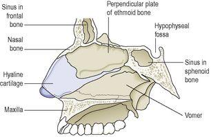
Figure 10.2
Structures forming the nasal septum.
The roof
is formed by the cribriform plate of the ethmoid bone and the sphenoid bone, frontal bone and nasal bones.
The floor
is formed by the roof of the mouth and consists of the hard palate in front and the soft palate behind. The hard palate is composed of the maxilla and palatine bones and the soft palate consists of involuntary muscle.
The medial wall
is formed by the septum.
The lateral walls
are formed by the maxilla, the ethmoid bone and the inferior conchae (
Fig. 10.3
).
Figure 10.3
Lateral wall of right nasal cavity.
The posterior wall
is formed by the posterior wall of the pharynx.
Lining of the nose
The nose is lined with very vascular
ciliated columnar epithelium
(ciliated mucous membrane,
Fig. 10.12
, respiratory mucosa) which contains mucus-secreting goblet cells (
p. 34
). At the anterior nares this blends with the skin and posteriorly it extends into the nasal part of the pharynx.
Openings into the nasal cavity
The anterior nares
, or nostrils, are the openings from the exterior into the nasal cavity. Nasal hairs are found here, coated in sticky mucus.
The posterior nares
are the openings from the nasal cavity into the pharynx.
The paranasal sinuses
are cavities in the bones of the face and the cranium, containing air. There are tiny openings between the paranasal sinuses and the nasal cavity. They are lined with mucous membrane, continuous with that of the nasal cavity. The main sinuses are:
•
maxillary sinuses in the lateral walls
•
frontal and sphenoidal sinuses in the roof (
Fig. 10.3
)
•
ethmoidal sinuses in the upper part of the lateral walls.
The sinuses function in speech and also lighten the skull.
The nasolacrimal ducts
extend from the lateral walls of the nose to the conjunctival sacs of the eye (
p. 199
). They drain tears from the eyes.
Respiratory function of the nose
The nose is the first of the respiratory passages through which the inspired air passes. The function of the nose is to begin the process by which the air is warmed, moistened and filtered.
The projecting
conchae
(
Figs 10.3
and
10.4
) increase the surface area and cause turbulence, spreading inspired air over the whole nasal surface. The large surface area maximises warming, humidification and filtering.
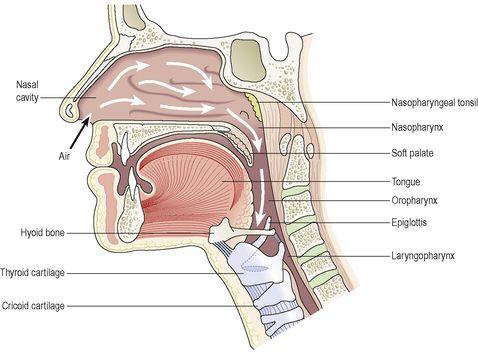
Figure 10.4
The pathway of air from the nose to the larynx.
Warming
This is due to the immense vascularity of the mucosa. This explains the large blood loss when a nosebleed (epistaxis) occurs.
Filtering and cleaning
Hairs at the anterior nares trap larger particles. Smaller particles such as dust and bacteria settle and adhere to the mucus. Mucus protects the underlying epithelium from irritation and prevents drying. Synchronous beating of the cilia wafts the mucus towards the throat where it is swallowed or coughed up (expectorated).
Humidification
As air travels over the moist mucosa, it becomes saturated with water vapour. Irritation of the nasal mucosa results in
sneezing
, a reflex action that forcibly expels an irritant.
The sense of smell
The nose is the organ of the sense of smell (
olfaction
). Nerve endings that detect smell are located in the roof of the nose in the area of the cribriform plate of the ethmoid bones and the superior conchae. These nerve endings are stimulated by airborne odours. The resultant nerve signals are carried by the
olfactory nerves
to the brain where the sensation of smell is perceived (
p. 200
).
Pharynx
Learning outcomes
After studying this section, you should be able to:
describe the location of the pharynx
relate the structure of the pharynx to its function.
Position
The pharynx is a tube 12 to 14 cm long that extends from the base of the skull to the level of the 6th cervical vertebra. It lies behind the nose, mouth and larynx and is wider at its upper end.
Structures associated with the pharynx
Superiorly
– the inferior surface of the base of the skull
Inferiorly
– it is continuous with the oesophagus
Anteriorly
– the wall is incomplete because of the openings into the nose, mouth and larynx
Posteriorly
– areolar tissue, involuntary muscle and the bodies of the first six cervical vertebrae.
For descriptive purposes the pharynx is divided into three parts:
nasopharynx, oropharynx
and
laryngopharynx
.
The nasopharynx
The nasal part of the pharynx lies behind the nose above the level of the soft palate. On its lateral walls are the two openings of the
auditory tubes
(
p. 187
), one leading to each middle ear. On the posterior wall are the
pharyngeal tonsils
(adenoids), consisting of lymphoid tissue. They are most prominent in children up to approximately 7 years of age. Thereafter they gradually atrophy.
The oropharynx
The oral part of the pharynx lies behind the mouth, extending from below the level of the soft palate to the level of the upper part of the body of the 3rd cervical vertebra. The lateral walls of the pharynx blend with the soft palate to form two folds on each side. Between each pair of folds is a collection of lymphoid tissue called the
palatine tonsil
.


