Ross & Wilson Anatomy and Physiology in Health and Illness (77 page)
Read Ross & Wilson Anatomy and Physiology in Health and Illness Online
Authors: Anne Waugh,Allison Grant
Tags: #Medical, #Nursing, #General, #Anatomy

The autonomic or involuntary part of the nervous system (
Fig. 7.1
) controls the ‘automatic’ functions of the body, i.e. those initiated in the brain below the level of the cerebrum. Although stimulation does not occur voluntarily, the individual may be conscious of its effects, e.g. an increase in their heart rate.
The effects of autonomic activity are rapid and the effector organs are:
•
smooth muscle, e.g. changes in airway or blood vessel diameter
•
cardiac muscle, e.g. changes in rate and force of the heartbeat
•
glands, e.g. increasing or decreasing gastrointestinal secretions.
The
efferent (motor) nerves
of the autonomic nervous system arise from the brain and emerge at various levels between the midbrain and the sacral region of the spinal cord. Many of them travel within the same nerve sheath as peripheral nerves to reach the organs that they innervate.
The autonomic nervous system is separated into two divisions:
•
sympathetic
(thoracolumbar outflow)
•
parasympathetic
(craniosacral outflow).
The two divisions have both structural and functional differences. They normally work in an opposing manner, thereby maintaining balance of involuntary functions. Sympathetic activity tends to predominate in stressful situations and parasympathetic activity during rest.
Each division has two efferent neurones between the central nervous system and effector organs. These are:
•
the preganglionic neurone
•
the postganglionic neurone.
The cell body of the preganglionic neurone is in the brain or spinal cord. Its axon terminals synapse with the cell body of the postganglionic neurone in an
autonomic ganglion
outside the CNS. The postganglionic neurone conducts impulses to the effector organ.
Sympathetic nervous system
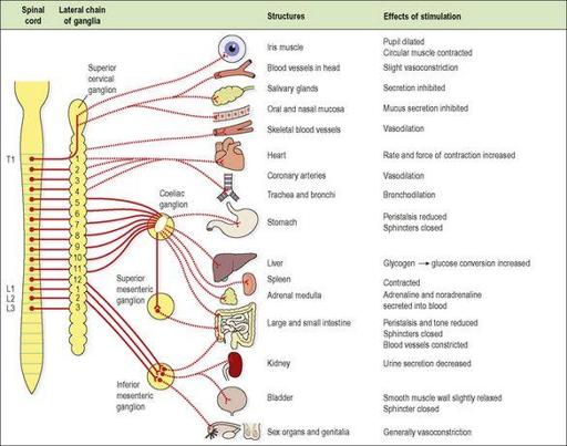
Since the preganglionic neurones originate in the spinal cord at the thoracic and lumbar levels, the alternative name of ‘thoracolumbar outflow’ is apt (
Fig. 7.46
).
Figure 7.46
The sympathetic outflow, the main structures supplied and the effects of stimulation.
Solid red lines – preganglionic fibres; broken lines – postganglionic fibres. There is a right and left lateral chain of ganglia.
The preganglionic neurone
This has its cell body in the
lateral column of grey matter
in the spinal cord between the levels of the 1st thoracic and 2nd or 3rd lumbar vertebrae. The nerve fibre of this cell leaves the cord by the anterior root and terminates at a synapse in one of the ganglia either in the
lateral chain of sympathetic ganglia
or passes through it to one of the
prevertebral ganglia
(see below). Acetylcholine is the neurotransmitter at sympathetic ganglia.
The postganglionic neurone
This has its cell body in a ganglion and terminates in the organ or tissue supplied. Noradrenaline (norepinephrine) is usually the neurotransmitter at sympathetic effector organs. The major exception is that there is no parasympathetic supply to the sweat glands, the skin and blood vessels of skeletal muscles. These structures are supplied by only sympathetic postganglionic neurones, which are known as sympathetic cholinergic nerves and usually have acetylcholine as their neurotransmitter (see
Fig. 7.8
).
Sympathetic ganglia
The lateral chains of sympathetic ganglia
These chains extend from the upper cervical level to the sacrum, one chain lying on each side of the vertebral bodies. The ganglia are attached to each other by nerve fibres. Preganglionic neurones that emerge from the cord may synapse with the cell body of the postganglionic neurone at the same level or they may pass up or down the chain through one or more ganglia before synapsing. For example, the nerve that dilates the pupil of the eye leaves the cord at the level of the 1st thoracic vertebra and passes up the chain to the superior cervical ganglion before it synapses with the cell body of the postsynaptic neurone. The postganglionic neurones then pass to the eyes.
The arrangement of the ganglia allows excitation of nerves at multiple levels very quickly, providing a rapid and widespread sympathetic response.
Prevertebral ganglia
There are three prevertebral ganglia situated in the abdominal cavity close to the origins of arteries of the same names:
•
coeliac ganglion
•
superior mesenteric ganglion
•
inferior mesenteric ganglion.
The ganglia consist of nerve cell bodies rather diffusely distributed among a network of nerve fibres that form plexuses. Preganglionic sympathetic fibres pass through the lateral chain to reach these ganglia.
Parasympathetic nervous system
Two neurones (preganglionic and postganglionic) are involved in the transmission of impulses from their source to the effector organ (
Fig. 7.47
). The neurotransmitter at both synapses is acetylcholine.
The preganglionic neurone
This is usually long in comparison to its counterpart in the sympathetic nervous system and has its cell body either in the brain or in the spinal cord. Those originating in the brain are the cranial nerves III, VII, IX and X, arising from nuclei in the midbrain and brain stem, and their nerve fibres terminate at or near effector organs. The cell bodies of the
sacral outflow
are in the lateral columns of grey matter at the distal end of the spinal cord. Their fibres leave the cord in sacral segments 2, 3 and 4 and synapse with postganglionic neurones in the walls of pelvic organs.
The postganglionic neurone
This is usually very short and has its cell body either in a ganglion or, more often, in the wall of the organ supplied.
Functions of the autonomic nervous system
The autonomic nervous system is involved in many complex involuntary reflex activities which, like the reflexes described previously, depend on sensory input to the brain or spinal cord, and on motor output. In this case the reflex action is rapid contraction, or inhibition of contraction, of involuntary (smooth and cardiac) muscle or glandular secretion. These activities are coordinated subconsciously in the brain, i.e. below the level of the cerebrum. Some sensory input does reach consciousness and may result in temporary inhibition of the reflex action, e.g. reflex micturition can be inhibited temporarily.
The majority of the body organs are supplied by both sympathetic and parasympathetic nerves, which have opposite effects that are finely balanced to ensure their optimum functioning meets body needs at any moment.
Sympathetic stimulation
prepares the body to deal with exciting and stressful situations, e.g. strengthening its defences in times of danger and in extremes of environmental temperature. A range of emotional states, e.g. fear, embarrassment and anger, also cause sympathetic stimulation. The adrenal glands are stimulated to secrete the hormones adrenaline (epinephrine) and noradrenaline (norepinephrine) into the bloodstream. These hormones potentiate and sustain the effects of sympathetic stimulation. It is said that sympathetic stimulation mobilises the body for ‘fight or flight’. The effects of stimulation on the heart, blood vessels and lungs (see below) enable the body to respond by preparing it for exercise. Additional effects are an increase in the metabolic rate and increased conversion of glycogen to glucose. During exercise, e.g. fighting or running away, when oxygen and energy requirements of skeletal muscles are greatly increased, these changes enable the body to respond quickly to meet the increased energy demand.
Parasympathetic stimulation
has a tendency to slow down body processes except digestion and absorption of food and the functions of the genitourinary systems. Its general effect is that of a ‘peace maker’, allowing restoration processes to occur quietly and peacefully.
Normally the two systems function together, maintaining a regular heartbeat, normal temperature and an internal environment compatible with the immediate external surroundings.
Effects of autonomic stimulation
Cardiovascular system
Sympathetic stimulation
•
Accelerates firing of the sinoatrial node in the heart, increasing the rate and force of the heartbeat.
•
Dilates the coronary arteries, increasing the blood supply to cardiac muscle.
•
Dilates the blood vessels supplying skeletal muscle, increasing the supply of oxygen and nutritional materials and the removal of metabolic waste products, thus increasing the capacity of the muscle to work.
•
Raises peripheral resistance and blood pressure by constricting the small arteries and arterioles in the skin. In this way an increased blood supply is available for highly active tissue, such as skeletal muscle, heart and brain.
•
Constricts the blood vessels in the secretory glands of the digestive system. This raises the volume of blood available for circulation in dilated blood vessels.
•
Accelerates blood coagulation because of vasoconstriction.
Parasympathetic stimulation
•
Decreases the rate and force of the heartbeat.
•
Constricts the coronary arteries, reducing the blood supply to cardiac muscle.
The parasympathetic nervous system exerts little or no effect on blood vessels except the coronary arteries.
Respiratory system
Sympathetic stimulation
This causes smooth muscle relaxation and therefore dilation of the airways (
bronchodilation
), especially the bronchioles, allowing a greater amount of air to enter the lungs at each inspiration, and increases the respiratory rate. In conjunction with the increased heart rate, the oxygen intake and carbon dioxide output of the body are increased to deal with ‘fight or flight’ situations.
Parasympathetic stimulation
This causes contraction of the smooth muscle in the airway walls, leading to
bronchoconstriction
.
Digestive and urinary systems
Sympathetic stimulation
•
The liver
increases conversion of glycogen to glucose, making more carbohydrate immediately available to provide energy.
•
The stomach
and
small intestine
. Smooth muscle contraction (peristalsis) and secretion of digestive juices are inhibited, delaying digestion, onward movement and absorption of food, and the tone of sphincter muscles is increased.
•
The adrenal (suprarenal) glands
are stimulated to secrete adrenaline (epinephrine) and noradrenaline (norepinephrine) which potentiate and sustain the effects of sympathetic stimulation throughout the body.
•
Urethral
and
anal sphincters
. The muscle tone of the sphincters is increased, inhibiting micturition and defecation.

