Ross & Wilson Anatomy and Physiology in Health and Illness (47 page)
Read Ross & Wilson Anatomy and Physiology in Health and Illness Online
Authors: Anne Waugh,Allison Grant
Tags: #Medical, #Nursing, #General, #Anatomy

The
ulnar artery
runs downwards on the ulnar or medial aspect of the forearm to cross the wrist and pass into the hand.
There are anastomoses between the radial and ulnar arteries, called the
deep
and
superficial palmar arches
, from which
palmar metacarpal
and
palmar digital arteries
arise to supply the structures in the hand and fingers.
Venous return from the upper limb
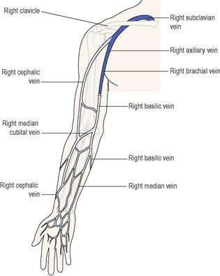
The veins of the upper limb are divided into two groups: deep and superficial veins (
Fig. 5.40
).
Figure 5.40
The main veins of the right arm
. Dark blue indicates deep veins.
The
deep veins
follow the course of the arteries and have the same names:
•
palmar metacarpal veins
•
deep palmar venous arch
•
ulnar and radial veins
•
brachial vein
•
axillary vein
•
subclavian vein.
The
superficial veins
begin in the hand and consist of the following:
•
cephalic vein
•
basilic vein
•
median vein
•
median cubital vein.
The
cephalic vein
begins at the back of the hand where it collects blood from a complex of superficial veins, many of which can be easily seen. It then winds round the radial side to the anterior aspect of the forearm. In front of the elbow it gives off a large branch, the
median cubital vein
, which slants upwards and medially to join the
basilic vein
. After crossing the elbow joint the cephalic vein passes up the lateral aspect of the arm and in front of the shoulder joint to end in the axillary vein. Throughout its length it receives blood from the superficial tissues on the lateral aspects of the hand, forearm and arm.
The
basilic vein
begins at the back of the hand on the ulnar aspect. It ascends on the medial side of the forearm and upper arm then joins the axillary vein. It receives blood from the medial aspect of the hand, forearm and arm. There are many small veins which link the cephalic and basilic veins.
The
median vein
is a small vein that is not always present. It begins at the palmar surface of the hand, ascends on the front of the forearm and ends in the basilic vein or the median cubital vein.
The
brachiocephalic vein
is formed when the subclavian and internal jugular veins unite. There is one on each side.
The
superior vena cava
is formed when the two brachiocephalic veins unite. It drains all the venous blood from the head, neck and upper limbs and terminates in the right atrium. It is about 7 cm long and passes downwards along the right border of the sternum.
Descending aorta in the thorax
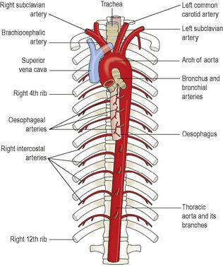
This part of the aorta is continuous with the arch of the aorta and begins at the level of the 4th thoracic vertebra. It extends downwards on the anterior surface of the bodies of the thoracic vertebrae (
Fig. 5.41
) to the level of the 12th thoracic vertebra, where it passes behind the diaphragm to become the abdominal aorta.
Figure 5.41
The aorta and its main branches in the thorax.
The descending aorta in the thorax gives off many
paired branches
which supply the walls of the thoracic cavity and the organs within the cavity, including the:
•
bronchial arteries
that supply the bronchi and their branches, connective tissue in the lungs and the lymph nodes at the root of the lungs
•
oesophageal arteries
, supplying the oesophagus
•
intercostal arteries
that run along the inferior border of the ribs and supply the intercostal muscles, some muscles of the thorax, the ribs, the skin and its underlying connective tissues.
Venous return from the thoracic cavity
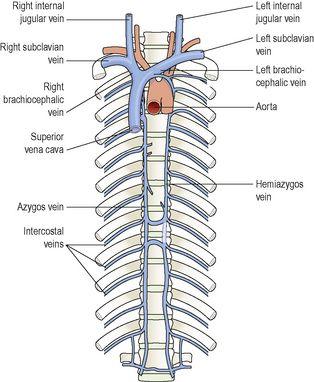
Most of the venous blood from the organs in the thoracic cavity is drained into the
azygos vein
and the
hemiazygos
vein (
Fig. 5.42
). Some of the main veins that join them are the
bronchial, oesophageal
and
intercostal veins
. The azygos vein joins the superior vena cava and the hemiazygos vein joins the left brachiocephalic vein. At the distal end of the oesophagus, some oesophageal veins join the azygos vein, and others the left gastric vein. A venous plexus is formed by anastomoses between the veins joining the azygos vein and those joining the left gastric veins, linking the general and portal circulations (see
Fig. 12.46, p. 313
).
Figure 5.42
The superior vena cava and the main veins of the thorax.
Abdominal aorta
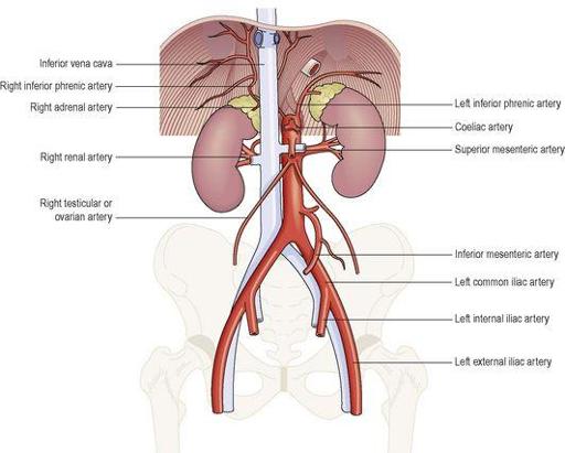
The abdominal aorta is a continuation of the thoracic aorta. The name changes when the aorta enters the abdominal cavity by passing behind the diaphragm at the level of the 12th thoracic vertebra. It descends in front of the bodies of the vertebrae to the level of the 4th lumbar vertebra, where it divides into the
right
and
left common iliac arteries
(
Fig. 5.43
).
Figure 5.43
The abdominal aorta and its branches.
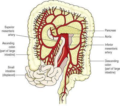
When a branch of the abdominal aorta supplies an organ it is only named here and is described in more detail in association with the organ. However, illustrations showing the distribution of blood from the coeliac, superior and inferior mesenteric arteries are presented here (
Figs 5.44
and
5.45
).
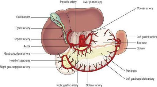
Figure 5.44
The coeliac artery and its branches, and the inferior phrenic arteries.
Figure 5.45
The superior and inferior mesenteric arteries and their branches.
Many branches arise from the abdominal aorta, some of which are paired and some unpaired.
Paired branches
•
Inferior phrenic arteries
supply the diaphragm.
•
Renal arteries
supply the kidneys and give off branches, the suprarenal arteries, to supply the adrenal glands.
•
Testicular arteries
supply the testes in the male.
•
Ovarian arteries
supply the ovaries in the female.
The testicular and ovarian arteries are much longer than the other paired branches, because these organs begin their development in the region of the kidneys. As they grow, they descend into the scrotum and the pelvis respectively, and are accompanied by their blood vessels.
Unpaired branches
The
coeliac artery
(
Fig. 5.43
) is a short thick artery about 1.25 cm long. It arises immediately below the diaphragm and divides into three branches:
•
the
left gastric artery
supplies the stomach
•
the
splenic artery
supplies the pancreas and the spleen
•
the
hepatic artery
supplies the liver, gall bladder and parts of the stomach, duodenum and pancreas.
The
superior mesenteric artery
(
Fig. 5.43
) branches from the aorta between the coeliac artery and the renal arteries. It supplies the whole of the small intestine and the proximal half of the large intestine.
The
inferior mesenteric artery
(
Fig. 5.43
) arises from the aorta about 4 cm above its division into the common iliac arteries. It supplies the distal half of the large intestine and part of the rectum.
Venous return from the abdominal organs
The
inferior vena cava
is formed when the
right
and
left common iliac veins
join at the level of the body of the 5th lumbar vertebra. This is the largest vein in the body, and it carries blood from all parts of the body below the diaphragm to the right atrium of the heart. It passes through the central tendon of the diaphragm at the level of the 8th thoracic vertebra.
