Ross & Wilson Anatomy and Physiology in Health and Illness (185 page)
Read Ross & Wilson Anatomy and Physiology in Health and Illness Online
Authors: Anne Waugh,Allison Grant
Tags: #Medical, #Nursing, #General, #Anatomy

Figure 16.33
The right humerus.
Anterior view.
The distal end of the bone presents two surfaces that articulate with the radius and ulna to form the elbow joint.
Ulna and radius (
Fig. 16.34
)
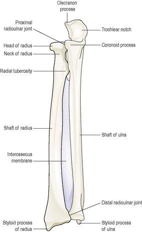
These are the two bones of the forearm. The ulna is longer than and medial to the radius and when the arm is in the anatomical position, i.e. with the palm of the hand facing forward, the two bones are parallel. They articulate with the humerus at the
elbow joint
, the carpal bones at the
wrist joint
and with each other at the
proximal
and
distal radioulnar
joints. In addition, an interosseous membrane, a fibrous joint, connects the bones along their shafts, stabilising their association and maintaining their relative positions despite forces applied from the elbow or wrist.
Figure 16.34
The right radius and ulna with the interosseous membrane
. Anterior view.
Carpal (wrist) bones (
Fig. 16.35
)
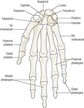
There are eight carpal bones arranged in two rows of four. From outside inwards they are:
•
proximal row
: scaphoid, lunate, triquetrum, pisiform
•
distal row
: trapezium, trapezoid, capitate, hamate.
Figure 16.35
The bones of the hand, wrist and fingers.
Anterior view.
These bones are closely fitted together and held in position by ligaments that allow a limited amount of movement between them. The bones of the proximal row are associated with the wrist joint and those of the distal row form joints with the metacarpal bones. Tendons of muscles lying in the forearm cross the wrist and are held close to the bones by strong fibrous bands, called retinacula (see
Fig. 16.50, p. 407
).
Metacarpal bones (bones of the hand)
These five bones form the palm of the hand. They are numbered from the thumb side inwards. The proximal ends articulate with the carpal bones and the distal ends with the phalanges.
Phalanges (finger bones)
There are 14 phalanges, three in each finger and two in the thumb. They articulate with the metacarpal bones and with each other, by hinge joints.
Pelvic girdle and lower limb
The lower limb forms a joint with the trunk at the pelvic girdle.
The pelvic girdle
The pelvic girdle is formed from two innominate (hip) bones. The
pelvis
is the term given to the basin-shaped structure formed by the pelvic girdle and its associated sacrum.
Innominate (hip) bones (
Fig. 16.36
)
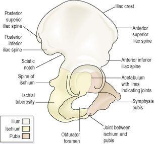
Each hip bone consists of three fused bones: the
ilium
,
ischium
and
pubis
. On its lateral surface is a deep depression, the
acetabulum
, which forms the hip joint with the almost-spherical head of femur.
Figure 16.36
The right hip bone.
Lateral view.
The
ilium
is the upper flattened part of the bone and it presents the
iliac crest
, the anterior curve of which is called the
anterior superior iliac spine
. The ilium forms a synovial joint with the sacrum, the
sacroiliac joint
, a strong joint capable of absorbing the stresses of weight bearing and which tends to become fibrosed in later life.
The
pubis
is the anterior part of the bone and it articulates with the pubis of the other hip bone at a cartilaginous joint, the
symphysis pubis
.
The
ischium
is the inferior and posterior part. The rough inferior projections of the ischia, the
ischial tuberosities
, bear the weight of the body when seated.
The union of the three parts takes place in the
acetabulum
.
The pelvis (
Fig. 16.37
)
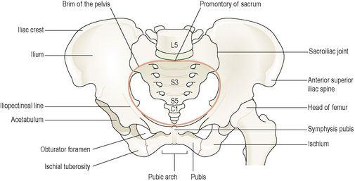
The pelvis is formed by the hip bones, the sacrum and the coccyx. It is divided into upper and lower parts by the
brim of the pelvis
, consisting of the promontory of the sacrum and the iliopectineal lines of the innominate bones. The
greater
or
false pelvis
is above the brim and the lesser or
true pelvis
is below.
Figure 16.37
The bones of the pelvis and the upper part of the left femur.
Differences between male and female pelves (
Fig. 16.38
)
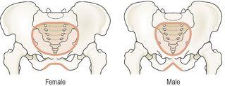
The shape of the female pelvis allows for the passage of the baby during childbirth. In comparison with the male pelvis, the female pelvis has lighter bones, is more shallow and rounded and is generally roomier.
Figure 16.38
The difference in shape of the male and female pelves.
The lower limb
Femur (thigh bone) (
Fig. 16.39
)
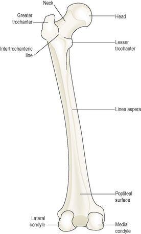
The femur is the longest and heaviest bone of the body. The head is almost spherical and fits into the
acetabulum
of the hip bone to form the
hip joint
. The neck extends outwards and slightly downwards from the head to the shaft and most of it is within the capsule of the hip joint.
Figure 16.39
The left femur.
Posterior view.
The posterior surface of the lower third forms a flat triangular area called the
popliteal surface
. The distal extremity has two articular
condyles
, which, with the tibia and patella, form the knee joint. The function of the femur is to transmit the weight of the body through the bones below the knee to the foot.
Tibia (shin bone) (
Fig. 16.40
)
The tibia is the medial of the two bones of the lower leg. The proximal extremity is broad and flat and presents two
condyles
for articulation with the femur at the
knee joint
. The head of the fibula articulates with the inferior aspect of the lateral condyle, forming the
proximal tibiofibular
joint.
Figure 16.40
The left tibia and fibula with the interosseous membrane.
Anterior view.
The distal extremity of the tibia forms the
ankle joint
with the talus and the fibula. The
medial malleolus
is a downward projection of bone medial to the ankle joint.
Fibula (
Fig. 16.40
)
