Ross & Wilson Anatomy and Physiology in Health and Illness (184 page)
Read Ross & Wilson Anatomy and Physiology in Health and Illness Online
Authors: Anne Waugh,Allison Grant
Tags: #Medical, #Nursing, #General, #Anatomy

Figure 16.29
The sternum and its attachments.
The
manubrium
is the uppermost section and articulates with the clavicles at the
sternoclavicular joints
and with the first two pairs of ribs.
The
body
or
middle portion
gives attachment to the ribs.
The
xiphoid process
is the tip of the bone. It gives attachment to the diaphragm, muscles of the anterior abdominal wall and the
linea alba
.
Ribs
The 12 pairs of ribs form the lateral walls of the thoracic cage (
Fig. 16.28
). They are elongated curved bones (
Fig. 16.30
) that articulate posteriorly with the vertebral column. Anteriorly, the first seven pairs of ribs articulate directly with the sternum and are known as the
true ribs
. The next three pairs articulate only indirectly. In both cases,
costal cartilages
attach the ribs to the sternum. The lowest two pairs of ribs, referred to as
floating ribs
, do not join the sternum at all, their anterior tips being free.
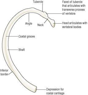
Figure 16.30
A typical rib
– viewed from below.
Each rib forms up to three joints with the vertebral column. Two of these joints are formed between facets on the head of the rib and facets on the bodies of two vertebrae, the one above the rib and the one below. Ten of the ribs also form joints between the tubercle of the rib and the transverse process of (usually) the lower vertebra.
The inferior surface of the rib is deeply grooved, providing a channel along which intercostal nerves and blood vessels run. Between each rib and the one below are the intercostal muscles, essential for breathing.
Because of the arrangement of the ribs, and the quantity of cartilage present in the ribcage, it is a flexible structure that can change its shape and size during breathing. The first rib is firmly fixed to the sternum and to the 1st thoracic vertebra, and does not move during inspiration. Because it is a fixed point, when the intercostal muscles contract, they pull the entire ribcage upwards towards the first rib. The mechanism of breathing is described on
page 247
.
Appendicular skeleton
Learning outcomes
After studying this section you should be able to:
identify the bones forming the appendicular skeleton
state the characteristics of the bones forming the appendicular skeleton
outline the differences in structure between the male and female pelves.
The appendicular skeleton consists of the shoulder girdle with the upper limbs and the pelvic girdle with the lower limbs (
Fig. 16.9
).
Shoulder girdle and upper limb
The upper limb forms a joint with the trunk via the shoulder (pectoral) girdle.
Shoulder girdle
The shoulder girdle consists of two scapulae and two clavicles.
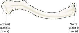
Clavicle (collar bone) (
Fig. 16.31
)
The clavicle is an S-shaped long bone. It articulates with the manubrium of the sternum at the
sternoclavicular joint
and forms the
acromioclavicular joint
with the
acromion process
of the scapula. The clavicle provides the only bony link between the upper limb and the axial skeleton.
Figure 16.31
The right clavicle.
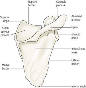
Scapula (shoulder blade) (
Fig. 16.32
)
The scapula is a flat triangular-shaped bone, lying on the posterior chest wall superficial to the ribs and separated from them by muscles.
Figure 16.32
The right scapula.
Posterior view.
At the lateral angle is a shallow articular surface, the
glenoid cavity
, which, with the
head of the humerus
, forms the
shoulder joint
.
On the posterior surface runs a rough ridge called the
spine
, which extends beyond the lateral border of the scapula and overhangs the glenoid cavity. The prominent overhang, which can be felt through the skin as the highest point of the shoulder, is called the
acromion process
and forms a joint with the clavicle, the
acromioclavicular joint
, a slightly movable synovial joint that contributes to the mobility of the shoulder girdle. The
coracoid process
, a projection from the upper border of the bone, gives attachment to muscles that move the shoulder joint.
The upper limb
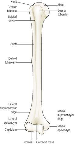
Humerus (
Fig. 16.33
)
This is the bone of the upper arm. The head sits within the glenoid cavity of the scapula, forming the shoulder joint. Distal to the head are two roughened projections of bone, the
greater
and
lesser tubercles
, and between them there is a deep groove, the
bicipital groove
or
intertubercular sulcus
, occupied by one of the tendons of the biceps muscle.


