Ross & Wilson Anatomy and Physiology in Health and Illness (131 page)
Read Ross & Wilson Anatomy and Physiology in Health and Illness Online
Authors: Anne Waugh,Allison Grant
Tags: #Medical, #Nursing, #General, #Anatomy

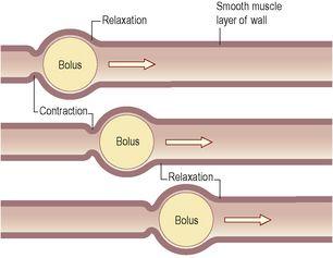
Contraction and relaxation of these muscle layers occurs in waves, which push the contents of the tract onwards. This type of contraction of smooth muscle is called
peristalsis
(
Fig. 12.4
) and is under the influence of sympathetic and parasympathetic nerves. Muscle contraction also mixes food with the digestive juices. Onward movement of the contents of the tract is controlled at various points by
sphincters
, which are thickened rings of circular muscle. Contraction of sphincters regulates forward movement. They also act as valves, preventing backflow in the tract. This control allows time for digestion and absorption to take place.
Figure 12.4
Movement of a bolus by peristalsis.
Submucosa
This layer consists of loose areolar connective tissue containing collagen and some elastic fibres, which binds the muscle layer to the mucosa. Within it are plexuses of blood vessels and nerves, lymph vessels and varying amounts of lymphoid tissue. The blood vessels are arterioles, venules and capillaries. The nerve plexus is the
submucosal plexus
(Meissner’s plexus,
Fig. 12.2
), containing sympathetic and parasympathetic nerves that supply the mucosal lining.
Mucosa
This consists of three layers of tissue:
•
mucous membrane
formed by columnar epithelium is the innermost layer, and has three main functions:
protection
,
secretion
and
absorption
•
lamina propria
consisting of loose connective tissue, which supports the blood vessels that nourish the inner epithelial layer, and varying amounts of lymphoid tissue that has a protective function
•
muscularis mucosa
, a thin outer layer of smooth muscle that provides involutions of the mucosa layer, e.g. gastric glands (
p. 291
), villi (
p. 294
).
Mucous membrane
In parts of the tract that are subject to great wear and tear or mechanical injury, this layer consists of
stratified squamous epithelium
with mucus-secreting glands just below the surface. In areas where the food is already soft and moist and where secretion of digestive juices and absorption occur, the mucous membrane consists of
columnar epithelial cells
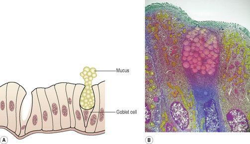
interspersed with mucus-secreting goblet cells (
Fig. 12.5
). Mucus lubricates the walls of the tract and protects them from the damaging effects of digestive enzymes. Below the surface in the regions lined with columnar epithelium are collections of specialised cells, or glands, which release their secretions into the lumen of the tract. The secretions include:
•
saliva
from the salivary glands
•
gastric juice
from the gastric glands
•
intestinal juice
from the intestinal glands
•
pancreatic juice
from the pancreas
•
bile
from the liver.
Figure 12.5
Columnar epithelium with a goblet cell. A.
Diagram.
B.
Coloured transmission electron micrograph of a section through a goblet cell (pink and blue) of the small intestine.
These are
digestive juices
and most contain enzymes that chemically break down food. Under the epithelial lining are varying amounts of lymphoid tissue that provide protection against ingested microbes.
Nerve supply
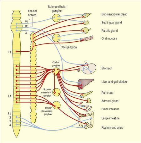
The alimentary canal and its related accessory organs are supplied by nerves from both divisions of the autonomic nervous system, i.e. both parasympathetic and sympathetic parts (
Fig. 12.6
). Their actions are antagonistic and one has a greater influence than the other, according to body needs, at any particular time. When digestion is required, this is normally through increased activity of the parasympathetic nervous system.
Figure 12.6
Autonomic nerve supply to the digestive system.
Parasympathetic – blue; sympathetic – red.
The parasympathetic supply
One pair of cranial nerves, the
vagus nerves
, supplies most of the alimentary canal and the accessory organs. Sacral nerves supply the most distal part of the tract. The effects of parasympathetic stimulation are:
•
increased muscular activity, especially peristalsis, through increased activity of the myenteric plexus
•
increased glandular secretion, through increased activity of the submucosal plexus (
Fig. 12.2
).
The sympathetic supply
This is provided by numerous nerves that emerge from the spinal cord in the thoracic and lumbar regions. These form plexuses (ganglia) in the thorax, abdomen and pelvis, from which nerves pass to the organs of the alimentary tract. The effects of sympathetic stimulation are to:
•
decrease muscular activity, especially peristalsis, because there is less stimulation of the myenteric plexus
•
decrease glandular secretion, as stimulation of the submucosal plexus is reduced.
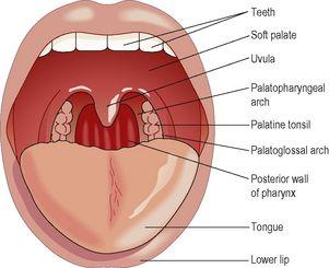
Mouth (
Fig. 12.7
)
Figure 12.7
Structures seen in the widely open mouth.
Learning outcomes
After studying this section, you should be able to:
list the principal structures associated with the mouth
describe the structure of the mouth
describe the structure and function of the tongue
describe the structure and function of the teeth


