Resurrecting Pompeii (30 page)
Read Resurrecting Pompeii Online
Authors: Estelle Lazer

Alveolar loss was dif ficult to assess in cases where teeth had not survived. Most of the cases that could be scored for both mandibles and maxillae showed some degree of interdental alveolar resorption. There were relatively few examples of considerable alveolar loss.
Bisel found that about 75 per cent of the Herculaneum sample exhibited some degree of alveolar resorption. Of these, 60 per cent had only slight resorption and 9 per cent displayed a moderate to marked degree of periodontal disease. Because of the post mortem loss of so many teeth it was not possible to adequately assess the degree of alveolar recession in the Pompeian sample. Nonetheless, the data that could be collected suggested the incidence and degree of alveolar resorption was comparable with that of the Herculaneum sample.
23
Abscesses are the result of the build-up of dental infections that can be causally related to caries, excessive, rapid attrition, trauma and periodontal disease. These can generally be detected as a clearly defined circular cavity in the alveolus near the root of the affected tooth. These cavities function as drains for the pus produced by the abscess. Bony changes to the alveolus that could be attributed to dental abscesses were recorded for each maxilla and mandible. It should be noted that deep pockets from advanced periodontal disease are virtually impossible to distinguish from abscesses.
24
There was a higher frequency of abscesses in the maxillae than the mandibles in the Pompeian sample, with at least one abscess in almost 43 per cent of the maxillae and just under 19 per cent of the mandibles. This is consistent with observations from other archaeological sites. A strong association was observed between advancing age and the presence and degree of abscesses in the skeletal series from Pueblo sites in Northeastern Arizona that were studied by Martin
et al
.
25
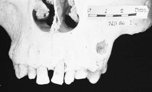
The Pompeian data was difficult to assess in this respect as it was not possible to estimate age-at-death in a number of cases where teeth and other age indicators had not survived. In the cases where assessment was possible, there did appear to be some correlation between age and the presence of dental abscesses, especially severe abscesses, which were almost always associated with older individuals (for example, Figure 8.1).
26
Bisel recorded a mean of 0.73 abscesses per male mouth and 0.66 per female mouth in the Herculanean sample. This is a higher frequency than that for the Pompeian sample. Capasso observed a total of 52 abscesses in 23 individuals in his sample, 51 of which were periapical. Nine of these individuals had only one abscess, eight had two, one had three, two individuals presented with four and two had five, while one individual had a total of eight oral abscesses. Capasso suggested that these abscesses were associated with the development of carious lesions and severe tooth attrition, which involved exposure of the pulp cavity.
27
Calculus is mineralized plaque. It is attached to the surface of the tooth. The mineral, though deposited from plaque fluid, derives from saliva. As a result the greatest amount of calculus formation occurs at sites on teeth that are nearest to the salivary glands. The reasons for plaque mineralization are not fully known, though it is thought that bacteria are involved. The presence and degree of calculus observed in the Pompeian sample was recorded.
28

Figure 8.1
Large sinus for abscess drainage (TF NS 86: 1). This abscess formed as a result of severe attrition (7.4), where tooth wear was so great that the pulp cavity was exposed to the air, making it susceptible to bacterial infection
Most of the surviving teeth of the Pompeian sample had, at least, a slight calculus deposit. It is important to note that it was only possible to score minimal expression as the storage conditions were not conducive to the preservation of larger deposits. The degree of calculus deposited on approximately half of the teeth for both mandibles and maxillae was recorded as slight. Only 19.5 per cent of the available maxillary and 11.1 per cent of the mandibular teeth displayed no evidence of calculus. This suggests that oral hygiene, as it is known to modern Western communities, was not a high priority.
Bisel did not present data on the presence of calculus on the Herculaneum teeth, whereas Capasso commented that there was a particularly high frequency of calculus deposits in the sample he studied. Forty-two, or 39 per cent, of the 139 mouths he studied showed some degree of calculus deposition. Twenty-eight individuals displayed slight; ten, medium; and four had considerable deposits of calculus.
29
The presence of linear enamel hypoplasia is the direct result of the failure of the enamel to properly form in the developing tooth. It can result from a variety of factors, such as nutritional stress, infection, poisoning or trauma to the tooth or pulp of a deciduous tooth lying over a growing permanent tooth. Enamel hypoplasia often presents as transverse line of indented enamel on the sides of the tooth crown, though it can also present as pits and grooves. They can only form during the period of crown development, which means that if all the dentition is examined, presence of hypoplasia can potentially reflect the health and nutrition of an individual between approximately the second and fourteenth year of life. The position of the line gives some indication of the age of the individual when such periods of stress occurred. It should be noted, however, that absence of hypoplasia does not necessarily infer that there were no periods of stress in the years of crown formation. While the presence of enamel disruption suggests insult, its absence cannot automatically be interpreted as evidence of a healthy and well-nourished person. Modern individuals with a history of lengthy and major illness in early childhood do not always display enamel hypoplasia.
30
Goodman and Armelagos discovered that anterior teeth were more susceptible to hypoplasias than posterior teeth because their developmental timing can be easily disrupted. Individuals exposed to the same environmental stressors may exhibit varying degrees of enamel hypoplasia. Martin
et al
. suggested that it was only really useful to record linear enamel hypoplasia in the case of anterior teeth.
31
Equipment was not available for making accurate readings for the distance between lines in the Pompeian sample, so no attempt was made to determine the period in juvenile tooth growth when the disruption occurred. Instead, the presence or absence of these lines was noted and a score was allocated with respect to the number of lines and degree of disruption to the enamel surface. Some degree of linear enamel hypoplasia was observed on 19 of the 33 maxillary teeth and 36 of the 45 mandibular teeth.
The Pompeian sample exhibited both a higher incidence and a greater degree of linear enamel hypoplasia on mandibular teeth. Nonetheless, most of the recorded cases of hypoplasia for the sample were slight. It is not reasonable to extrapolate interpretations from the small Pompeian sample onto the population at large, beyond the observation that a number of individuals experienced episodes of stress, possibly as a result of illness, during the period of crown formation.
In the posthumous publication of Bisel ’s work, the frequency of linear enamel hypoplasia is recorded at about 50 per cent. Her earlier publications indicate lower levels, with observations of some degree of linear enamel hypoplasia on about 25 per cent of the male and 16 per cent of the female Herculanean sample. Capasso observed some degree of enamel hypoplasia in the teeth of 71 individuals in the sample he studied. Of these, 42 were male and 29 were identified as female, which meant a male to female ratio of approximately 3 : 2. Petrone
et al
. examined the mouths of 51 individuals and recorded a frequency of enamel hypoplasia in 96.1 per cent, with some degree of expression in 36 males and 15 females.
32
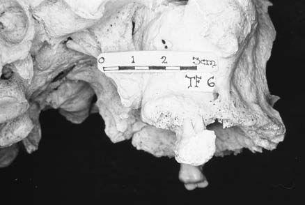
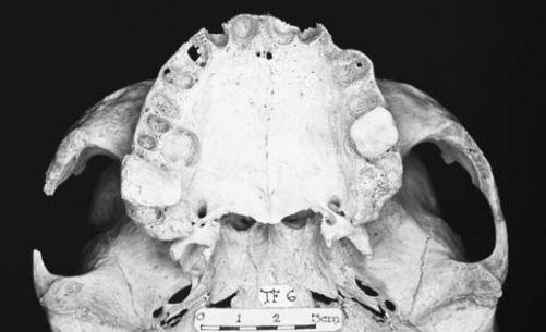
No unequivocal evidence of dental intervention was observed in the available Pompeian sample. It is possible that teeth were extracted ante mortem, though it is impossible to distinguish intentional from unintentional ante mortem tooth loss from the skeletal evidence. There is some indication that tooth extraction was not practised even when removal might have alleviated a problem. A good example of this can be seen in the maxilla of a skull in the Forum Baths collection (see Figures 8.2 and 8.3).
33
Only two teeth are still
in situ
, the right second molar and the left first molar. The former tooth is marked by a very thick deposit of calculus over the entire tooth, with the exception of a small polished facet on the mesial side of the crown. This wear facet is not on the normal occlusal surface. Considerable alveolar loss is apparent, with the exposure of more than half the roots of the remaining molar. The left first molar displays no sign of calculus. The degree of alveolar loss is comparable to that on the right side of the maxilla.
The excessive build-up of calculus on the right molar suggested that this individual was avoiding the use of this side of the mouth for mastication. The apparent reason for this was a large abscess associated with the palatal root of the adjacent first molar. This involved most of the lingual alveolar surface as evidenced by the marked resorption of the palatal process of the alveolar region. There is also a small sinus from an abscess associated with the distal buccal root of this tooth (Figure 8.2). It appears that this molar
 Figure 8.2
Lateral view of skull showing excessive build-up of calculus on tooth adjacent to an abscessed tooth (TF 6)
 Figure 8.3
View of maxilla from below showing excessive build-up of calculus on tooth adjacent to an abscessed tooth (TF 6)
34
Non-dental evidence for dental practice
It is likely that, at least, some of the observed ante mortem tooth loss was the result of deliberate extraction, even though no tooth forceps have been found amongst the repertoire of medical instruments discovered in Pompeii. General bone forceps may have been substituted for this purpose. Other examples of tooth forceps found in Roman archaeological contexts were made of iron and it is remotely possible that this material did not survive burial in Pompeii.
35
There were specialists in extraction in the Roman world as can be seen in the case of a shop built into the podium of the Temple of Castor and Pollux in the Roman Forum, where dental procedures were undertaken, along with the sale of pharmaceutical and cosmetic goods. It was suggested that a combined dental and pharmaceutical practice would have been valuable as related problems could have been treated simultaneously. A total of 86 decayed teeth, including two molars from children, were found during excavation. There is evidence that they were removed by skilled practitioners as great care had been taken to ensure that the entire tooth was excised and that no portion of the roots remained in the sockets. In one case, a portion of alveolar bone was extracted with the tooth so that the extremely decayed crown did not break from the root. The dangers of leaving dental fragments in the jaw were discussed by Celsus in the sections on dentistry in his treatise,
De Medicina
, which he wrote in Italy in the early part of the first century
AD
. It is notable that he considered extraction to be a last resort, after the failure of other measures, like treatment of the gums. He also recommended the use of gold wire to bind teeth that had been loosened, by an accident or a blow, to firm teeth in order to stabilize them.
36
