Pocket Medicine: The Massachusetts General Hospital Handbook of Internal Medicine (132 page)
Read Pocket Medicine: The Massachusetts General Hospital Handbook of Internal Medicine Online
Authors: Marc Sabatine
Tags: #Medical, #Internal Medicine

BOOK: Pocket Medicine: The Massachusetts General Hospital Handbook of Internal Medicine
8.52Mb size Format: txt, pdf, ePub
1
Parasternal long-axis view
allows visualization of the right ventricle (RV), ventricular septum (VS), posterior wall (PW) aortic valve cusps, left ventricle (LV), mitral valve, left atrium (LA), and ascending thoracic aorta (Ao). *Pulmonary artery. (Top: From
Mayo Clinic Proceedings
. [Tajik AJ, Seward JB, Hagler DJ,
et al.
Two-dimensional real-time ultrasonic imaging of the heart and great vessels: Technique, image orientation, structure identification, and validation.
Mayo Clinic Proceedings
, 1978;53:271–303], with permission. Bottom: From Oh JK, Seward JB, Tajik AJ.
The Echo Manual
,
3rd ed
. Philadelphia: Lippincott Williams & Wilkins, 2006. By permission of Mayo Foundation for Medical Education and Research. All rights reserved.)
2
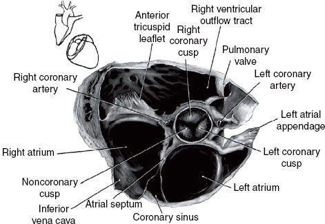
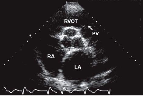
Parasternal short-axis view at the level of the aorta:
LA, left atrium; PV, pul-monary valve; RA, right atrium; RVOT, right ventricular outflow tract. (Top: From
Mayo Clinic Proceedings
. [Tajik AJ, Seward JB, Hagler DJ,
et al.
Two-dimensional real-time ultrasonic imaging of the heart and great vessels: Technique, image orientation, structure identification, and validation.
Mayo Clinic Proceedings
, 1978;53:271–303], with permission. Bottom: From Oh JK, Seward JB, Tajik AJ.
The Echo Manual
,
3rd ed
. Philadelphia: Lippincott Williams & Wilkins, 2006. By permission of Mayo Foundation for Medical Education and Research. All rights reserved.)
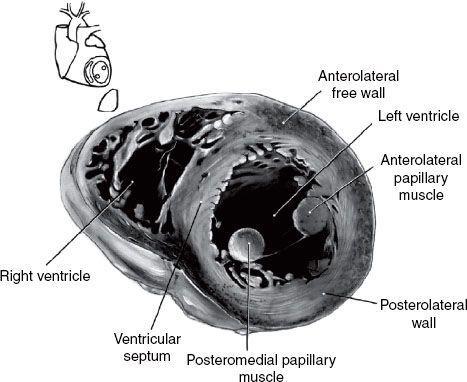
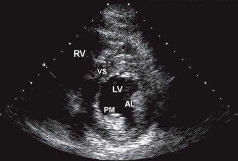
3 Parasternal short-axis view at the level of the papillary muscles:
AL, anterolateral papillary muscle; PM, posteromedial papillary muscle; RV, right ventricle; VS, ventricular septum; LV, left ventricle. (Top: From
Mayo Clinic Proceedings
. [Tajik AJ, Seward JB, Hagler DJ,
et al.
Two-dimensional real-time ultrasonic imaging of the heart and great vessels: Technique, image orientation, structure identification, and validation.
Mayo Clinic Proceedings
, 1978;53:271–303], with permission. Bottom: From Oh JK, Seward JB, Tajik AJ.
The Echo Manual
,
3rd ed
. Philadelphia: Lippincott Williams & Wilkins, 2006. By permission of Mayo Foundation for Medical Education and Research. All rights reserved.)
4
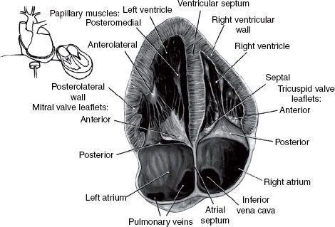
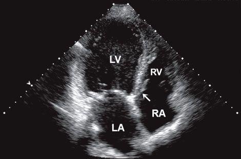
Apical four-chamber view:
Note that at some institutions the image is re-versed so that the left side of the heart appears on the right side of the screen. LA, left atrium; LV, left ventricle; RA, right atrium; RV, right ventricle. (Top: From
Mayo Clinic Proceedings
. [Tajik AJ, Seward JB, Hagler DJ,
et al.
Two-dimensional real-time ultrasonic imaging of the heart and great vessels: Technique, image orientation, structure identification, and validation.
Mayo Clinic Proceedings
, 1978;53:271–303], with permission. Bottom: From Oh JK, Seward JB, Tajik AJ.
The Echo Manual
,
3rd ed
. Philadelphia: Lippincott Williams & Wilkins, 2006. By permission of Mayo Foundation for Medical Education and Research. All rights reserved.)
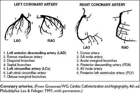
Coronary Angiography
Peripheral Blood Smears

1
Normal smear.
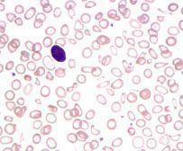
2
Hypochromic, microcytic anemia due to iron-deficiency.
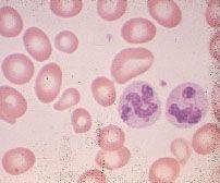
3
Macrocytic anemia due to pernicious anemia; note macro-ovalocytes and hypersegmented neutrophils.

Other books
Run Away Home by Terri Farley
Night & Demons by David Drake
Delicious Desires by Jackie Williams
The Last Song of Orpheus by Robert Silverberg
Andanzas y malandanzas by Alberto Rivas Bonilla
Gone at Zero Hundred 00:00 by Cr Hiatt
A Woman Gone Mad by Kimber S. Dawn
Cartilage and Skin by Michael James Rizza
Kelly's Chance by Brunstetter, Wanda E.
Cooper Security 06 - Secret Intentions by Paula Graves