Pediatric Examination and Board Review (88 page)
Read Pediatric Examination and Board Review Online
Authors: Robert Daum,Jason Canel

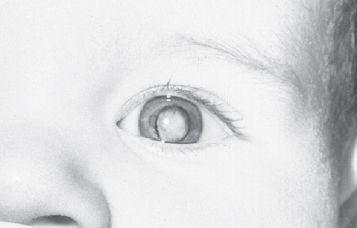
FIGURE 54-1.
Leukocoria of the left eye caused by retrolental membrane (persistent hyperplastic primary vitreous or persistent fetal vasculature). (Reproduced, with permission, from Hay Jr WW, Levin MJ, Sondheimer JM, et al. Current Diagnosis & Treatment: Pediatrics, 19th ed. New York: McGraw-Hill; 2009: Fig. 15-1.)
10.
(A)
11.
(C)
12.
(B)
Patients with Down syndrome can have epicanthal folds and Brushfield spots but not leukocoria. Cataracts are the most common cause of leukocoria in children, and all the other answers represent the differential diagnosis. About 55% of infants with congenital cataracts have a positive family history.
13.
(E)
Trisomy 13 (Patau syndrome), trisomy 18 (Edward syndrome), deficiency of galactose-1- uridyltransferase (galactosemia), and cri-du-chat syndrome (deletion of the short arm of chromosome 5) are all associated with cataracts in infancy. Children with these genetic diseases require regular ophthalmologic evaluation. The cataracts associated with galactosemia can be prevented by appropriate diet.
14.
(B)
Of patients with sporadic aniridia (see next answer), one-fifth will develop Wilms tumor. Thus these patients require renal ultrasound as a screening tool every 3-6 months until they are 5 years old. Also, aniridia leads to glaucoma in 75% of patients.
15.
(B)
Two-thirds of cases are autosomal dominant; one-third are sporadic. The gene for aniridia has been localized to 11p13. Aniridia is bilateral in 98% (see
Figure 54-2
).
FIGURE 54-2.
Bilateral aniridia. Iris remnants present temporally in each eye. (Reproduced, with permission, from Hay Jr WW, Levin MJ, Sondheimer JM, et al. Current Diagnosis & Treatment: Pediatrics, 19th ed. New York: McGraw-Hill; 2009: Fig. 15-21.)
16.
(C)
CHARGE syndrome (coloboma, heart disease, atresia choanae, retarded growth and development, genital anomalies, and ear anomalies), the trisomies, and nevus sebaceous are all associated with colobomas. VATER association (vertebral defects, anal atresia, tracheoesophageal fistula, radial dysplasia, renal anomaly) is not (see
Figure 54-3
).
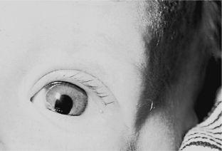
FIGURE 54-3.
Iris coloboma located inferiorly. (Reproduced, with permission, from Hay Jr WW, Levin MJ, Sondheimer JM, et al. Current Diagnosis & Treatment: Pediatrics, 19th ed. New York: McGraw-Hill; 2009: Fig. 15-20.)
17.
(B)
Blood in the anterior chamber almost always represents a hyphema in children. Tumors and coagulopathies can present similarly but are rare (see
Figure 54-4
).
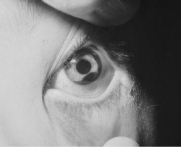
FIGURE 54-4.
Hyphema filling approximately 20% of the anterior chamber. (Reproduced, with permission, from Hay Jr WW, Levin MJ, Sondheimer JM, et al. Current Diagnosis & Treatment: Pediatrics, 19th ed. New York: McGraw-Hill; 2009: Fig. 15-10.)
18.
(D)
Treatment for hyphema is supportive with bed rest and head elevation to 30-45 degrees to promote resorption. Steroids, both topical and oral, have been used as have topical mydriatics. Surgical drainage is not a recommended treatment (see
Figure 54-5
).
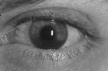
FIGURE 54-5.
Hyphema. This hyphema is just beginning to layer out reflecting its acute nature. (Reproduced, with permission, from Knoop KJ, Stack LB, Storrow AS, et al. Atlas of Emergency Medicine, 3rd ed. New York: McGraw-Hill; 2010:82. Photo contributor: Lawrence B. Stack, MD.)
19.
(C)
Rebleeding most often occurs 3-5 days after the initial bleed and can lead to other complications. The treatments discussed above are aimed at preventing rebleeding.
 S
S
UGGESTED
R
EADING
American Academy of Ophthalmology Web site.
www.aao.org
. Accessed June 2010.
Beck AD. Diagnosis and management of pediatric glaucoma.
Ophthalmol Clin North Am.
2001;14(3):501-512.
Fallaha N. Pediatric cataracts.
Ophthalmol Clin North Am.
2001;14(3):479-492.
Michael JG. Management of corneal abrasion in children: a randomized clinical trial.
Ann Emerg Med.
2002;40(1):67-72.
Wilson SA, Last A. Management of corneal abrasions.
Am Fam
Physician.
2004;70(1):123-8.
CASE 55: A 5-YEAR-OLD BOY WITH A PAINLESS LIMP
A 5-year-old boy has come to your office today because his mother has noticed him limping on the right for 3-4 days. He does not recall any trauma to the area and has not been ill in the last month. He has had no fevers and denies any pain in the back, hip, knee, or ankle.
On examination he is afebrile. His weight is 18 kg (50%), height is 39.5 inches (<5%), and body mass index (BMI) is 18.3 (>95%). Upon walking across the examination room, you note an obvious limp on the right. Examination of the leg reveals no swelling, erythema, or warmth. You note that he is holding the leg in external rotation and has discomfort and decreased range of motion on internal rotation and abduction. His knee and ankle examinations are normal.
SELECT THE ONE BEST ANSWER
1.
What is the most likely diagnosis?
(A) transient synovitis
(B) SCFE (slipped capital femoral epiphysis)
(C) Legg-Calvé-Perthes disease
(D) septic arthritis
(E) growing pains
2.
What is the cause of this disorder?