Pediatric Examination and Board Review (90 page)
Read Pediatric Examination and Board Review Online
Authors: Robert Daum,Jason Canel

FIGURE 55-2.
Slipped capital femoral epiphysis. Anteroposterior (AP) and Frog-leg views of a slipped epiphysis. The dotted lines show the normal position of the femoral head. (Reproduced, with permission, from Skinner HB. Current Diagnosis & Treatment in Orthopedics, 4th ed. New York: McGraw-Hill; 2006: Fig. 11-13.)
8.
(E)
SCFE is more common in obese or rapidly growing pubertal boys. It is bilateral 20-50% of the time. Each side usually presents at different times.
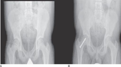
FIGURE 55-3.
Radiographic images of a young patient with a slipped capital femoral epiphysis, before (A) and after (B) screw fixation. (Reproduced, with permission, from Brunicardi FC, Andersen DK, Billiar TR, et al. Schwartz’s Principles of Surgery, 9th ed. New York: McGraw-Hill; 2010: Fig. 43-57A,B.)
9.
(C)
Because further slippage of the capital femoral epiphysis will occur without treatment, surgical correction is necessary. The most popular technique is pinning through the femoral head and neck to stabilize the area (see
Figure 55-3
).
10.
(B)
Osgood-Schlatter disease is a common cause of knee pain in athletic adolescents. Males are affected more than females. It manifests as pain and sometimes swelling over the tibial tubercle that is made worse by activities that involve pressure with bending of the knee: squatting, jumping, and kneeling (see
Figure 55-4
).
11.
(B)
12.
(C)
NSAIDs, ice, and rest as needed are the preferred and common treatments of Osgood-Schlatter disease. Steroid injections are not advised, and treatment is almost always nonsurgical. Braces offer minimal support.
13.
(E)
When noting more than a mildly abnormal Adams test (forward bending to assess for posterior chest asymmetry, the screening tool for scoliosis) in a premenarchal girl, radiographic evaluation is an appropriate first step to assess the degree of severity of the scoliosis. The risk for progression of scoliosis is much greater in premenarchal girls and should be pursued more aggressively. Posterior to anterior radiographs of the thorax should be ordered to determine the Cobb angle of deviation (the angle made by the intersection of 2 lines drawn parallel to the uppermost and lowermost vertebrae involved in the curve) (see
Figure 55-5
). Incidentally, PA radiographs subject the breast tissue to less radiation than anterior to posterior films.
14.
(E)
Twenty percent of patients with congenital scoliosis have genitourinary defects; 15% have congenital heart disease. Other skeletal malformations are also common.
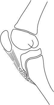
FIGURE 55-4.
Osgood-Schlatter disease. There is pain and swelling over the tibial tubercle. The radiographs would show characteristic fragmentation of the tibial tubercle apophysis, similar to diagram. (Reproduced, with permission, from Skinner HB. Current Diagnosis & Treatment in Orthopedics, 4th ed. New York: McGraw-Hill; 2006: Fig. 11-29.)
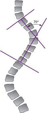
FIGURE 55-5.
Cobb method. Use of Cobb method to measure the scoliotic curve. First lines are drawn along the endplates of the upper and lower vertebrae that are maximally tilted into the concavity of the curve. Next, a perpendicular line is drawn to each of the earlier-drawn lines. The angle of intersection is the Cobb angle.
15.
(C)
Screening examinations should start at 6-7 years old when girls are premenarchal because the risk of progression is highest at this time. Although in 2004 the USPSTF (United States Preventive Services Task Force) released a statement recommending against routine screening for idiopathic scoliosis in adolescents, the AAP (American Academy of Pediatrics) and AAOS (American Academy of Orthopedic Surgeons) take the opposite view, recommending routine screening for all prepubertal children and adolescents (see
Figure 55-6
).
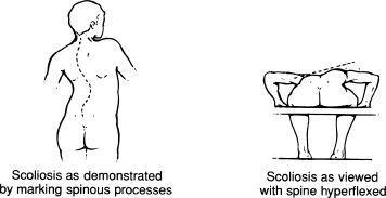
FIGURE 55-6.
Structural scoliosis. Inspection of the flexed spine from behind shows the unequal elevation of the two erector spinae muscle masses. (Reproduced, with permission, from LeBlond RF, DeGowin RL, Brown DD. DeGowin’s Diagnostic Examination, 9th ed. New York: McGraw-Hill; 2009: Fig. 13-13.)
16.
(D)
An angle of 60 degrees or more is severe scoliosis and often leads to cardiopulmonary sequelae. When the angle is 25 degrees or more, observation is recommended. Between 25 and 45 degrees, close follow-up, physical therapy, and occasionally a brace are sufficient treatment. When 45 degrees or more, surgical treatment is often required.
17.
(E)
18.
(E)
Scheuermann kyphosis is a clinical entity distinct from postural kyphosis, which has a clearly understood cause and can be corrected by the patient during the examination. On radiographs of the back, Scheuermann kyphosis has typical findings of wedging of 3 or more thoracic vertebrae and loss of the anterior height of the affected vertebrae. Patients with this disorder, when mild, require no treatment. But unlike postural kyphosis, many require brace and exercise programs (see
Figure 55-7
).
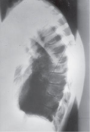
FIGURE 55-7.
Scheuermann kyphosis is characterized by vertebral wedging endplate changes and kyphosis. (Reproduced, with permission, from Skinner HB. Current Diagnosis & Treatmen)t in Orthopedics, 4th ed. New York: McGraw-Hill; 2006: Fig. 11-34.
 S
S
UGGESTED
R
EADING
Behrman RE, Kliegman RM, Jenson HB, et al.
Nelson Textbook of Pediatrics
. Philadelphia, PA: WB Saunders; 2007.
DeLee JC, Drez D Jr.
DeLee & Drez’s Orthopaedic Sports Medicine Principles and Practice
. Philadelphia, PA: WB Saunders; 2003:1831-1835.
Kim MK. The limping child.
Clin Pediatr Emerg Med.
2002; 3(2):129-137.
Richards BS, Vitale MG. Screening for idiopathic scoliosis in adolescents.
J Bone Joint Surg.
2008;90-A(1):195-198.
Sassmannshausen G. Back pain in the young athlete.
Clin Sports
Med.
2002;21(1):121-132.
CASE 56: A 4-YEAR-OLD WITH EAR PAIN
