Connectome (24 page)
Authors: Sebastian Seung

 Â
Â
Â
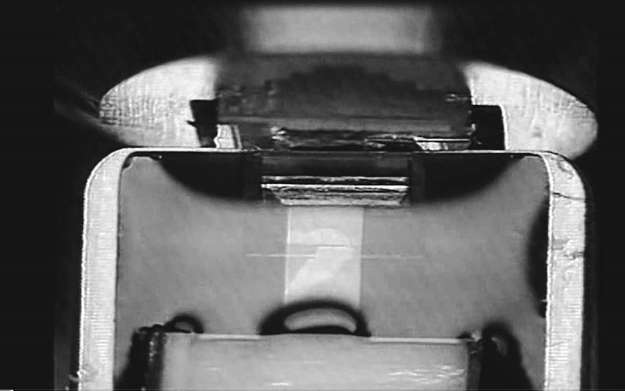
Figure 31. Freshly cut brain slices being collected by a plastic tape rising out of the water
Â
The first prototype of ATUM, the automated tape-collecting ultramicrotome, was built in more modest surroundingsâa garage thousands of miles away in the city of Alhambra, near Los Angeles. Its inventor, Ken Hayworth, is tall, thin, and bespectacled, with a determined walk and an intense way of talking. As an engineer at NASA's Jet Propulsion Laboratory, Hayworth built inertial guidance systems for spacecraft. Then he switched careers, enrolling in a neuroscience Ph.D. program at the University of Southern California. Hayworth has a lot of energy, which may explain why he used his spare time to build a new machine for slicing brains in his garage.
The prototype sliced at 10 microns, too thick for electron microscopy, but it demonstrated the basic idea. One day Hayworth received a phone call from out of the blue. It was Jeff Lichtman, the Harvard expert on synapse elimination, calling to suggest a collaboration. Hayworth set up shop at Harvard, and built another ATUM that was capable of cutting at 50 nanometers, the thickness achieved by a conventional ultramicrotome. Lichtman egged him on, and the machine eventually achieved 30 nanometers.
To image the slices, Hayworth teamed up with Narayanan “Bobby” Kasthuri. The two made an entertainingly odd couple. Other lab members joked that Kasthuri
seemed
crazy, with his wild hair and even wilder stories, but that Hayworth was actually the crazy one. (More on this inside joke later.) They and another researcher, Richard Schalek, employed a scanning electron microscope for imaging, the same instrument modified by Denk.
Denk's invention eliminates the need to collect sections; Hayworth's makes collecting them reliable. Other inventors are working on their own schemes to improve cutting and imaging. For example, Graham Knott has shown how to use a beam of ions to vaporize the top few nanometers off the top of the block. This technique is similar to Denk's but eliminates the need for a diamond knife.
Such inventions are just the beginning of what I anticipate will be a golden age in serial electron microscopy.
Along with that golden age comes a new challenge for neuroscience, the era of too much information. Just one cubic millimeter of brain tissue could yield a petabyte of image data. This is equivalent to a digital photo album containing one billion images. An entire mouse brain is a thousand times larger, a human brain a thousand times larger still. So the improvements in cutting, collecting, and imaging are not by themselves enough to find connectomes. Imaging every neuron and synapse will produce torrents of information, overwhelming the ability of any human to comprehend. To find connectomes, we need not only machines for
making
images, but also machines for
seeing
them.
The ancient greeks told the story of King Minos, who kept a beautiful white bull for himself instead of offering it as a sacrifice. The gods, angry at his greed, punished Minos by driving his wife mad with lust for the bull. She gave birth to the Minotaur, a monster with two legs and two horns. Minos imprisoned her deadly offspring in the Labyrinth, a mazelike structure ingeniously constructed by the great engineer Daedalus. Eventually the hero Theseus came from Athens and killed the Minotaur. To find his way back out of the Labyrinth, he followed a thread supplied by his lover Ariadne, the daughter of Minos.
Connectomics reminds me of this myth. Like the Labyrinth, the brain must deal with the consequences of destructive emotions such as greed and lust, while also inspiring acts of ingenuity and love. Try to imagine yourself traveling through the axons and dendrites of the brain, like Theseus navigating the twisting passages of the Labyrinth. Perhaps you are a protein molecule sitting on a molecular motor car running on a molecular track. You are being transported on the long journey from your birthplace, the cell body, to your destination, the outer reaches of the axon. You patiently sit and watch as the walls of the axon go by.
If this journey sounds intriguing, let me invite you to embark on a virtual version. You will travel through images of the brain, rather than the brain itself. You'll trace the path of an axon or dendrite through a stack of images collected by the machines described in Chapter 8. It's a task essential for finding connectomes. In order to map the brain's connections, you have to see which neurons are connected by synapses, and you can't do it without knowing where the “wires” go.
To find an entire connectome, though, you'd have to explore every passage in the brain's labyrinth. To map just one cubic millimeter, you'd have to travel through miles of neurites and wade through a petabyte of images. Such laborious and careful analysis would be essential; a mere glance at the images would tell you nothing. This style of science seems far removed from Galileo's sighting of the moons of Jupiter or Leeuwenhoek's glimpse of sperm.
Today, our notion of “science as seeing” is being stretched to the limit by current technologies. No single person can possibly comprehend all the images now being collected by automated instruments. But if technology created the problem, maybe it can also solve it. Perhaps computers could trace the paths of all those axons and dendrites through the images. If our machines did most of the work for us, we'd be able to see connectomes.
The problem of dealing with huge quantities of data is not unique to connectomics. The world's largest scientific project is the Large Hadron Collider (LHC), a circular tube constructed one hundred meters underground, inside a twenty-seven-kilometer-long tunnel between Lake Geneva and the Jura Mountains. The LHC accelerates protons to great speeds and smashes them together to probe the forces between elementary particles. At one location on its circumference sits a gigantic apparatus called the Compact Muon Solenoid. It's designed to detect one billion collisions
per second, of which one hundred are selected by computers that automatically sift through the data. Only these interesting events are recorded, but the data still flows at a torrential rate, as each event yields over one megabyte. The data is shipped to a network of supercomputers around the world for analysis.
To find entire connectomes of mammalian brains, we will need microscopes that produce images at data rates greater than those of the LHC. Can we analyze the data quickly enough to keep up? The scientists who compiled the
C. elegans
connectome encountered a similar challenge. To their surprise, it took more effort to analyze the images than to collect them.
Â
In the mid-1960s, the South African biologist Sydney Brenner saw the possibility of using serial electron microscopy to map all the connections in a small nervous system. The term
connectome
had not been invented yet,
and Brenner called the task “reconstruction of a nervous system.” Brenner was working at the MRC Laboratory for Molecular Biology in Cambridge, England. At that time, he and others at the lab were establishing
C. elegans
as a standard animal for research on genetics. It later became the first animal to have its genome sequenced, and thousands of biologists study
C. elegans
today.
Brenner thought that
C. elegans
might also help us understand the biological basis of behavior. It did the standard things like feeding, mating, and laying eggs. It also gave canned responses to certain stimuli. For example, if you touched its head, it would recoil and swim away. Now suppose you found a worm that was incapable of one of these standard behaviors. If its offspring inherited the same problem, you could assume that the cause was a genetic defect, and try to pinpoint it. That kind of research would elucidate the relationship between genes and behaviors, which would already be valuable. But one could raise the stakes even further by examining the nervous systems of such mutant worms. Perhaps one would be able to identify particular neurons or pathways disrupted by the faulty gene. The prospect of studying the worm at all these levelsâgenes, neurons, and behaviorâsounded truly exciting. But the whole plan hinged on something that Brenner did not have: a map of the normal worm's nervous system. Without that, it would be difficult to discern what was different about the nervous systems of mutants.
Brenner was aware of the early twentieth-century attempt of Richard Goldschmidt,
a German-American biologist, to map the nervous system of another species of worm,
Ascaris lumbricoides.
Goldschmidt's light microscope did not have enough resolution to show the branches of neurons clearly, or reveal synapses. Brenner decided to try something similar with
C. elegans,
but using the superior technology of the electron microscope and the ultramicrotome.
C. elegans
is just one millimeter long, much smaller than
Ascaris,
which can grow up to a foot in the intestines of its human hosts. Converting the entire
C. elegans
worm, like a tiny sausage, into slices thin enough for electron microscopy could be accomplished with a mere several thousand cuts. Nichol Thomson, a member of Brenner's team, found it impossible to slice up an entire worm without error, owing to the technical difficulties of the not-yet-automated slicing process, but he could manage a large fraction of a worm. Brenner decided to combine images from segments of several worms. It was a reasonable strategy because the worm's nervous system is so standardized.
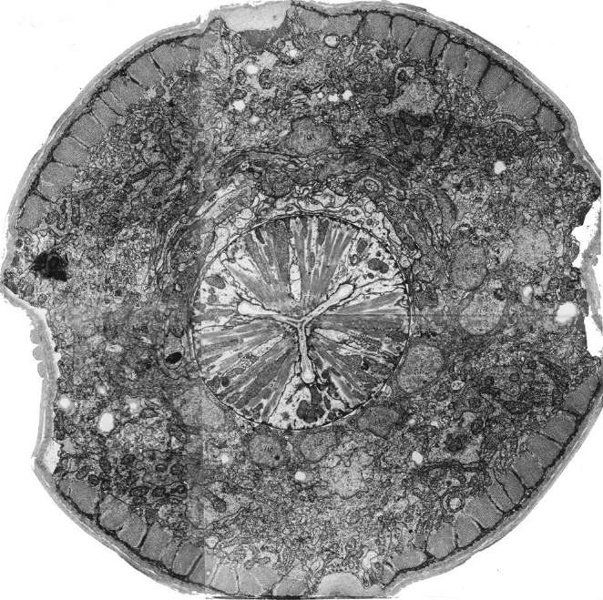
Thomson sliced up worms until he had covered every region of the worm's body at least once. The slices were placed one by one in an electron microscope and imaged (see Figure 32). This laborious process eventually yielded a stack of images representing the entire nervous system of
C. elegans
. All of the worm's synapses were there.
Â
 Â
Â
Â
Figure 32. A slice of C. elegans
Â
You might think Brenner and his team were done at that point. Isn't a connectome just the entirety of all synapses? In fact, they had only just begun. Although the synapses were all visible, their organization was still hidden. In effect, the researchers had collected a jumbled-up bag of synapses. To find the connectome, they needed to sort out which synapses belonged to which neurons. They couldn't tell from a single image, which showed only two-dimensional cross-sections of neurons. But if they could follow the successive cross-sections of a single neuron through a sequence of images, they could determine which synapses belonged to it. And if this could be done for all the neurons, then the connectome would be found. In other words, Brenner's team would know which neurons were connected to which other neurons.
Again, think of a worm as a tiny sausage. But imagine this time that the sausage is stuffed with spaghetti.
These spaghetti strands are its neurons, and our task is to trace the path of each one. Since we don't have x-ray vision, we ask the butcher to cut the sausage into many thin slices. Then we lay all the slices flat and trace each strand by matching its cut pieces from slice to slice.
To have any hope of tracing without errors, the slices must be extremely thin, less than the diameter of a spaghetti strand. Similarly, the slices of
C. elegans
had to be thinner than the branches of neurons, which can be less than 100 nanometers in diameter. Nichol Thomson cut slices about 50 nanometers thickâjust thin enough
to allow most branches of neurons to be traced reliably.
John White, who was trained as an electrical engineer, attempted to computerize the analysis of the images, but the technology was too primitive. White and a technician named Eileen Southgate had to resort to manual analysis. Cross-sections of the same neuron were marked with the same number or letter, as shown in the two images in Figure 33. To trace a single neuron in its entirety, the researchers repeatedly wrote the same symbol
on the appropriate cross-section in successive images, like Theseus unrolling Ariadne's thread in the Labyrinth. Once the paths of neurons were traced, they went back to each synapse and noted the letters or numbers of the neurons involved in it. And in this way the
C. elegans
connectome slowly emerged.