Ross & Wilson Anatomy and Physiology in Health and Illness (98 page)
Read Ross & Wilson Anatomy and Physiology in Health and Illness Online
Authors: Anne Waugh,Allison Grant
Tags: #Medical, #Nursing, #General, #Anatomy

Figure 9.7
The microscopic structure of the thyroid gland.
Thyroxine and tri-iodothyronine
Iodine is essential for the formation of the thyroid hormones, thyroxine (T
4
) and tri-iodothyronine (T
3
), so numbered as these molecules contain four and three atoms of the element iodine respectively. The body’s main dietary sources of iodine are seafood, vegetables grown in iodine-rich soil and iodinated table salt. The thyroid gland selectively takes up iodine from the blood, a process called iodine trapping.
The thyroid hormones are synthesised as large precursor molecules called
thyroglobulin
, the major constituent of colloid. The release of T
3
and T
4
into the blood is stimulated by
thyroid stimulating hormone
(TSH) from the anterior pituitary.
Secretion of TSH is stimulated by
thyrotrophin releasing hormone
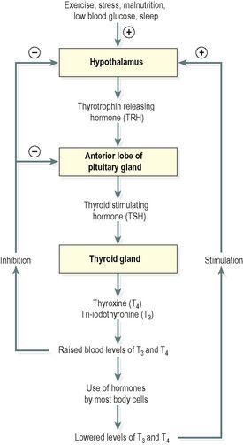
(TRH) from the hypothalamus and secretion of TRH is stimulated by exercise, stress, malnutrition, low plasma glucose levels and sleep. The level of secretion of TSH depends on the plasma levels of T
3
and T
4
because these hormones affect the sensitivity of the anterior pituitary to TRH. Through the negative feedback mechanism, increased levels of T
3
and T
4
decrease TSH secretion and vice versa (
Fig. 9.8
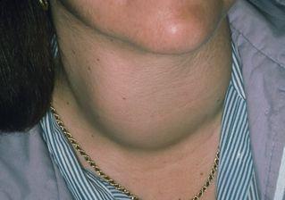
). When the supply of iodine is deficient, excess TSH is secreted and there is proliferation of thyroid gland cells and enlargement of the gland (goitre, see
Fig. 9.16
). Secretion of T
3
and T
4
begins about the third month of fetal life and is increased at puberty and in women during the reproductive years, especially during pregnancy. Otherwise, it remains fairly constant throughout life.
Figure 9.8
Negative feedback regulation of the secretion of thyroxine (T
4
) and tri-iodothyronine (T
3
).
Figure 9.16
Enlarged thyroid gland in goitre.
Thyroid hormones enter the cell nucleus and regulate gene expression, i.e. they increase or decrease the synthesis of some proteins including enzymes. They enhance the effects of other hormones, e.g. adrenaline (epinephrine) and noradrenaline (norepinephrine).
T
3
and T
4
affect most cells of the body by:
•
increasing the basal metabolic rate and heat production
•
regulating metabolism of carbohydrates, proteins and fats.
T
3
and T
4
are essential for normal growth and development, especially of the skeleton and nervous system. Most other organs and systems are also influenced by thyroid hormones – physiological effects of T
3
and T
4
on the heart, skeletal muscles, skin, digestive and reproductive systems are more evident when there is underactivity or overactivity of the thyroid gland. These changes are listed in
Table 9.3
and the effects, especially in childhood, can be profound.
Table 9.3
Common effects of abnormal secretion of thyroid hormones
| Hyperthyroidism: increased T 3 and T 4 secretion | Hypothyroidism: decreased T 3 and T 4 secretion |
|---|---|
| Increased basal metabolic rate | Decreased basal metabolic rate |
| Weight loss, good appetite | Weight gain, anorexia |
| Anxiety, physical restlessness, mental excitability | Depression, psychosis, mental slowness, lethargy |
| Hair loss | Dry skin, brittle hair |
| Tachycardia, palpitations, atrial fibrillation | Bradycardia |
| Warm sweaty skin, heat intolerance | Dry cold skin, prone to hypothermia |
| Diarrhoea | Constipation |
| Exophthalmos in Graves’ disease ( Fig. 9.15 ) |
Calcitonin
This hormone is secreted by the parafollicular or C-cells in the thyroid gland (
Fig. 9.7
). It acts on bone cells and the kidneys to reduce blood calcium (Ca
2+
) levels when they are raised. It promotes storage of calcium in bones and inhibits reabsorption of calcium by the renal tubules. Its effect is opposite to that of parathyroid hormone, the hormone secreted by the parathyroid glands. Release of calcitonin is stimulated by an increase in the blood calcium levels.
This hormone is important during childhood when bones undergo considerable changes in size and shape.
Parathyroid glands
Learning outcomes
After studying this section you should be able to:
describe the position and gross structure of the parathyroid glands
outline the functions of parathyroid hormone and calcitonin
explain how blood levels of parathyroid hormone and calcitonin are regulated.


