Ross & Wilson Anatomy and Physiology in Health and Illness (65 page)
Read Ross & Wilson Anatomy and Physiology in Health and Illness Online
Authors: Anne Waugh,Allison Grant
Tags: #Medical, #Nursing, #General, #Anatomy

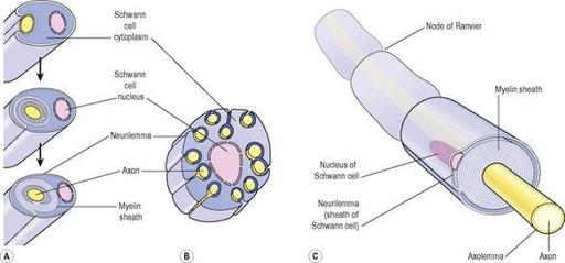
Figure 7.3
Arrangement of myelin
.
A.
Myelinated neurone.
B.
Non-myelinated neurone.
C.
Length of myelinated axon.
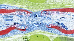
Figure 7.4
Node of Ranvier.
A colour transmission electron micrograph of a longitudinal section of a myelinated nerve fibre. Nerve tissue is shown in blue and myelin in red.
Non-myelinated neurones
Postganglionic fibres and some small fibres in the central nervous system are
non-myelinated
. In this type a number of axons are embedded in Schwann cell plasma membranes (
Fig. 7.3B
). The adjacent Schwann cells are in close association and there is no exposed axolemma. The speed of transmission of nerve impulses is significantly slower in non-myelinated fibres.
Dendrites
These are the many short processes that receive and carry incoming impulses towards cell bodies. They have the same structure as axons but are usually shorter and branching. In motor neurones dendrites form part of synapses (see
Fig. 7.7
) and in sensory neurones they form the sensory receptors that respond to specific stimuli.
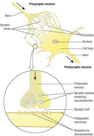
Figure 7.7
Diagram of a synapse.
The nerve impulse (action potential)
An impulse is initiated by stimulation of sensory nerve endings or by the passage of an impulse from another nerve. Transmission of the impulse, or action potential, is due to movement of ions across the nerve cell membrane. In the resting state the nerve cell membrane is polarised due to differences in the concentrations of ions across the plasma membrane. This means that there is a different electrical charge on each side of the membrane, which is called the
resting membrane potential
. At rest the charge on the outside is positive and inside it is negative. The principal ions involved are:
•
sodium (Na
+
), the main extracellular cation
•
potassium (K
+
), the main intracellular cation.
In the resting state there is a continual tendency for these ions to diffuse along their concentration gradients, i.e. K
+
outwards and Na
+
into cells. When stimulated, the permeability of the nerve cell membrane to these ions changes. Initially Na
+
floods into the neurone from the extracellular fluid causing
depolarisation
, creating a
nerve impulse
or
action potential
. Depolarisation is very rapid, enabling the conduction of a nerve impulse along the entire length of a neurone in a few milliseconds (ms). It passes from the point of stimulation in one direction only, i.e. away from the point of stimulation towards the area of resting potential. The one-way direction of transmission is ensured because following depolarisation it takes time for
repolarisation
to occur.
Almost immediately following the entry of sodium, K
+
floods out of the neurone and the movement of these ions returns the membrane potential to its resting state. This is called the
refractory period
during which restimulation is not possible. As the neurone returns to its original resting state, the action of the
sodium–potassium pump
expels Na
+
from the cell in exchange for K
+
(see
p. 33
).
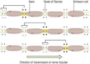
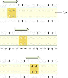
In myelinated neurones, the insulating properties of the myelin sheath prevent the movement of ions. Therefore electrical changes across the membrane can only occur at the gaps in the myelin sheath, i.e. at the nodes of Ranvier (see
Fig. 7.2
). When an impulse occurs at one node, depolarisation passes along the myelin sheath to the next node so that the flow of current appears to ‘leap’ from one node to the next. This is called
saltatory conduction
(
Fig. 7.5
).
Figure 7.5
Saltatory conduction of an impulse in a myelinated nerve fibre.
The speed of conduction depends on the diameter of the neurone: the larger the diameter, the faster the conduction. Myelinated fibres conduct impulses faster than unmyelinated fibres because saltatory conduction is faster than complete conduction, or
simple propagation
(
Fig. 7.6
). The fastest fibres can conduct impulses to, e.g., skeletal muscles at a rate of 130 metres per second while the slowest impulses travel at 0.5 metres per second.
Figure 7.6
Simple propagation of an impulse in a non-myelinated nerve fibre.
Arrows indicate the direction of impulse transmission.
The synapse and neurotransmitters
There is always more than one neurone involved in the transmission of a nerve impulse from its origin to its destination, whether it is sensory or motor. There is no physical contact between these neurones. The point at which the nerve impulse passes from one to another is the
synapse
(
Fig. 7.7
). At its free end, the axon of the
presynaptic neurone
breaks up into minute branches that terminate in small swellings called
synaptic knobs
, or terminal boutons. These are in close proximity to the dendrites and the cell body of the
postsynaptic neurone
. The space between them is the
synaptic cleft
. Synaptic knobs contain spherical
synaptic vesicles
, which store a chemical, the
neurotransmitter
that is released into the synaptic cleft. Neurotransmitters are synthesised by nerve cells, actively transported along the axons and stored in the synaptic vesicles. They are released by exocytosis in response to the action potential and diffuse across the synaptic cleft. They act on specific receptor sites on the postsynaptic membrane. Their action is short lived, because immediately they have acted upon the postsynaptic neurone or effector organ, such as a muscle fibre, they are either inactivated by enzymes or taken back into the synaptic knob. Knowledge of the action of the common neurotransmitters is important because some drugs mimic, neutralise (antagonise) or prolong their effect. Usually neurotransmitters have an excitatory effect at the synapse but they are sometimes inhibitory.
The neurotransmitters in the brain and spinal cord include
noradrenaline (norepinephrine), adrenaline (epinephrine), dopamine, histamine, serotonin, gamma aminobutyric acid (GABA)
and
acetylcholine
. Other substances, such as enkephalins, endorphins and substance P, have specialised roles in, for example, transmission of pain signals.
Figure 7.8
summarises the neurotransmitters of the peripheral nervous system.
