Ross & Wilson Anatomy and Physiology in Health and Illness (68 page)
Read Ross & Wilson Anatomy and Physiology in Health and Illness Online
Authors: Anne Waugh,Allison Grant
Tags: #Medical, #Nursing, #General, #Anatomy

Figure 7.16
The positions of the ventricles of the brain (in yellow) superimposed on its surface.
Viewed from the left side.
The lateral ventricles
These cavities lie within the cerebral hemispheres, one on each side of the median plane just below the corpus callosum. They are separated from each other by a thin membrane, the septum lucidum, and are lined with ciliated epithelium. They communicate with the third ventricle by
interventricular foramina
.
The third ventricle
The third ventricle is a cavity situated below the lateral ventricles between the two parts of the thalamus. It communicates with the fourth ventricle by a canal, the
cerebral aqueduct
.
The fourth ventricle
The fourth ventricle is a diamond-shaped cavity situated below and behind the third ventricle, between the
cerebellum
and
pons
. It is continuous below with the
central canal
of the spinal cord and communicates with the subarachnoid space by foramina in its roof. Cerebrospinal fluid enters the subarachnoid space through these openings and through the open distal end of the central canal of the spinal cord.
Cerebrospinal fluid (CSF)
Cerebrospinal fluid is secreted into each ventricle of the brain by
choroid plexuses
. These are vascular areas where there is a proliferation of blood vessels surrounded by ependymal cells in the lining of ventricle walls. CSF passes back into the blood through tiny diverticula of arachnoid mater, called
arachnoid villi
(arachnoid granulations,
Fig. 7.17
), which project into the venous sinuses. The movement of CSF from the subarachnoid space to venous sinuses depends upon the difference in pressure on each side of the walls of the arachnoid villi, which act as one-way valves. When CSF pressure is higher than venous pressure, CSF passes into the blood and when the venous pressure is higher the arachnoid villi collapse, preventing the passage of blood constituents into the CSF. There may also be some reabsorption of CSF by cells in the walls of the ventricles.
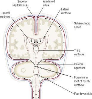
Figure 7.17
Arrows showing the flow of cerebrospinal fluid.
From the roof of the fourth ventricle CSF flows through foramina into the subarachnoid space and completely surrounds the brain and spinal cord (
Fig. 7.17
). There is no intrinsic system of CSF circulation but its movement is aided by pulsating blood vessels, respiration and changes of posture.
CSF is secreted continuously at a rate of about 0.5 ml per minute, i.e. 720 ml per day. The volume remains fairly constant at about 150 ml, as absorption keeps pace with secretion. CSF pressure may be measured using a vertical tube attached to a
lumbar puncture
needle inserted into the subarachnoid space above or below the 4th lumbar vertebra (which is below the end of the spinal cord). The pressure remains fairly constant at about 10 cm H
2
O when the individual is lying on his side and about 30 cm H
2
O when sitting up. If the brain is enlarged by, e.g., haemorrhage or tumour, some compensation is made by a reduction in the amount of CSF. When the volume of brain tissue is reduced, such as in degeneration or atrophy, the volume of CSF is increased. CSF is a clear, slightly alkaline fluid with a specific gravity of 1.005, consisting of:
•
water
•
mineral salts
•
glucose
•
plasma proteins: small amounts of albumin and globulin

•
a few leukocytes.
Functions of cerebrospinal fluid
CSF supports and protects the brain and spinal cord by maintaining a uniform pressure around these vital structures and acting as a cushion or shock absorber between the brain and the skull.
It keeps the brain and spinal cord moist and there may be exchange of nutrients and waste products between CSF and nerve cells. CSF is thought to be involved in regulation of breathing as it bathes the surface of the medulla where the central respiratory chemoreceptors are located (
Ch. 10
).
Brain
Learning outcomes
After studying this section you should be able to:
describe the blood supply to the brain
name the lobes and principal sulci of the brain
outline the functions of the cerebrum
identify the main sensory and motor areas of the cerebrum
outline the position and functions of the thalamus and hypothalamus
describe the position and functions of the midbrain, pons, medulla oblongata and reticular activating system
describe the structure and functions of the cerebellum.


