Pediatric Examination and Board Review (184 page)
Read Pediatric Examination and Board Review Online
Authors: Robert Daum,Jason Canel

(B)
Microsporum canis
(C)
Trichophyton tonsurans
(D)
Epidermophyton floccosum
(E)
Trichophyton rubrum
7.
An 8-year-old boy develops fever, headache, cough, and dyspnea. A chest radiograph reveals diffuse interstitial infiltrates. He is treated with oral antibiotics (amoxicillin-clavulanate) for 1 week without improvement. Fever and headache persist. He then becomes somnolent and confused. A spinal tap is performed that shows a high protein concentration, low glucose concentration, and pleocytosis, mostly lymphocytes. The diagnosis of cryptococcal meningitis is confirmed by cryptococcal antigen testing of the CSF. The boy likely has an underlying disorder involving
(A) humoral immunity
(B) cell-mediated immunity
(C) complement deficiency
(D) phagocytic function of neutrophils
(E) combined cell-mediated and humoral immunity
8.
A 6-month-old infant boy develops progressive respiratory distress with tachypnea and intercostal retractions but no fever. Bilateral rales are heard on auscultation. The infant has hypoxia with an oxygen saturation of 89% and requires intubation. The diagnosis of
P jiroveci
is confirmed by a fluoresceinconjugated monoclonal antibody stain of a tracheal aspirate specimen. All of the following drugs can be used alone for treatment of
P jiroveci
except
(A) atovaquone
(B) clindamycin
(C) pentamidine isethionate
(D) trimetrexate-leucovorin
(E) trimethoprim-sulfamethoxazole
9.
A 3-year-old girl who lives in a large urban city develops fever to 102°F (38.8°C), irritability, and drowsiness in a 2-week period. The child then develops vomiting, lethargy, and nuchal rigidity. A CT scan of the brain shows hydrocephalus and a ventriculoperitoneal shunt is placed. The CSF shows a cell count of 500/mm
3
with a lymphocyte predominance. The protein concentration was 85 mg/dL and the glucose level was 40 mg/dL. A chest radiograph is normal, and the Mantoux test is nonreactive. The child’s mother reveals that she and her daughter have been living in a homeless shelter for the past 6 months. The most likely diagnosis is
(A) cryptococcal meningitis
(B) neurocysticercosis
(C) Baylisascaris meningoencephalitis
(D) lymphocytic choriomeningitis
(E) tuberculous meningitis
10.
A full-term newborn infant boy is born to a 23-yearold mother who has a positive Mantoux test reaction, a result obtained 1 week before delivery. The infant should be evaluated for congenital tuberculosis in all of the following circumstances except when the
(A) mother has miliary tuberculosis
(B) mother has tuberculous endometritis
(C) mother has tuberculous infection of the knee joint
(D) mother has apical scarring of the right upper lobe of the lung with a negative sputum smear for acid-fast bacilli
(E) mother has active pulmonary tuberculosis
11.
A 4-year-old boy with HIV infection has a CD4 count of 80 cells/mm
3
(3%). He has a fever of 102°F (38.8°C) that persists for 1 week and is associated with weight loss, abdominal pain, fatigue, and diarrhea. Anemia is present with a hemoglobin of 9.0 g/dL. The most likely infectious etiology to explain these findings is
(A) nontuberculous mycobacteria, disseminated
(B)
Bartonella henselae
, disseminated
(C)
M tuberculosis
, disseminated
(D) brucellosis, disseminated
(E) histoplasmosis, disseminated
12.
A 3-year-old girl has a 1-month history of bilateral swelling of the submandibular lymph nodes. There is no history of fever, fatigue, or weight loss. A chest radiograph is normal and a Mantoux test result is positive at 10 mm. All of the following are consistent with nontuberculous mycobacterial lymphadenitis except
(A) age of the patient
(B) involvement of bilateral lymph nodes
(C) normal chest radiograph
(D) size of Mantoux test response
(E) lack of systemic symptoms
13.
A 2-year-old boy who attends a child-care center develops diarrhea that persists for 2 weeks and is associated with malaise, anorexia, abdominal distension, and abdominal cramps. You suspect giardiasis as the etiology. All of the following methods can be used to diagnose
Giardia lamblia
infection except
(A) duodenal fluid specimen
(B) concentrated specimen of stool
(C) DFA test of stool
(D) IgG-specific serum antibody to
Giardia
by enzyme immunoassay
(E) enzyme immunoassay to detect stool antigen
14.
Toxoplasmosis can cause both congenital infection and acquired infection in older children and adults. The most common physical finding in children with acquired toxoplasmosis is
(A) splenomegaly
(B) pharyngitis
(C) conjunctivitis
(D) macular rash
(E) lymphadenopathy
15.
A 16-year-old sexually active adolescent female presents to your office with a 1-week history of vaginal discharge and dysuria. She also reports a recent history of dyspareunia. A pelvic examination reveals vaginal and vulvar erythema with a frothy discharge. The cervix has a “strawberry appearance,” related to friability and punctate hemorrhages. Based on these findings, you strongly suspect infection with
(A)
Chlamydia trachomatis
(B)
Gardnerella vaginalis
(C)
Neisseria gonorrhoeae
(D)
Trichomonas vaginalis
(E)
Haemophilus ducreyi
16.
A 3-year-old boy develops fever and malaise in a 10-day period. On physical examination he has a fever of 102°F (38.8°C) and hepatosplenomegaly. The leukocyte count is 9000/mm
3
with 50% eosinophils (4500/mm
3
). An important epidemiologic finding to support the diagnosis of visceral larva migrans (VLM) caused by
Toxocara canis
is
(A) recent visit to a cave
(B) recent play in a sandbox
(C) recent play in a barn
(D) family has two adult dogs as pets
(E) consumption of raw or undercooked meat
17.
The different
Plasmodium
species that cause malaria have unique geographic distribution and resistance patterns. The following statement regarding the clinical features and epidemiology of malaria is correct
(A)
P ovale
infections are common only in South Africa
(B)
P vivax
malaria is prevalent on the Indian subcontinent and in Central and South America
(C)
P malariae
has the same geographic distribution as
P falciparum
and is more common in most areas they coinhabit
(D) relapses can occur in
P malariae
infection because of a persistent hepatic stage of infection
(E) most deaths worldwide from malaria occur in adolescents
18.
A 4-year-old girl returns from a trip to Bangladesh with her family where they traveled for the past 3 months. She develops diarrhea that persists for 2 weeks before being brought to your office by her parents. There is a history of bloody stools associated with abdominal pain. You suspect intestinal amebiasis. An important extraintestinal complication of
Entamoeba histolytica
infection you should consider is
(A) ameboma
(B) liver abscess
(C) brain abscess
(D) cutaneous amebiasis
(E) genitourinary tract involvement
ANSWERS
1.
(A)
The skin lesions of blastomycosis usually begin as papules that evolve into verrucous, ulcerative lesions (
Figure 104-1
). Osteomyelitis is the third most common manifestation of disseminated blastomycosis after lung and skin involvement. The CNS can also be involved with meningitis the most common complication.
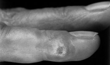
FIGURE 104-1.
Typical skin lesion of sporotrichosis involving the finger. (Reproduced, with permission, from Wolff K, Johnson RA. Fitzpatrick’s Color Atlas & Synopsis of Clinical Dermatology, 5th ed. New York: McGraw-Hill; 2005: 739.)
2.
(B)
Areas of high incidence of histoplasmosis include the Mississippi and Ohio River Valleys. Some activities of children that can predispose to infection include playing in barns, hollow trees, caves, or bird roosts. Infection is usually selflimited in immunocompetent children, but one form of infection called progressive disseminated histoplasmosis can occur. Prolonged fever, failure to thrive, and hepatosplenomegaly can develop in infants. A chest radiograph can show diffuse reticulonodular infiltrates. This form of the disease is often fatal if untreated.
3.
(E)
In children, sporotrichosis is localized to the skin and subcutaneous tissue (see
Figure 104-1
). The disease most commonly affects the face and extremities, in particular the hands and fingers. In most cases, infection will spread to the regional lymph nodes that drain the primary site of infection. Skin lesions associated with tuberculosis include erythema nodosum; papulonecrotic tuberculids that are miliary lesions of the skin, verrucosa cutis, which is a tuberculous wart-like lesion; and scrofuloderma, which is an ulcer or sinus tract resulting from rupture of a lymph node.
4.
(D)
The travel history and the clinical situation suggest that the patient has coccidioidomycosis. The infection is endemic to Southern California, Arizona, western and southern Texas, and New Mexico. In children with coccidioidomycosis, an acute diffuse erythematous rash and erythema multiforme are common. The primary infection involves the lungs, and, in healthy children, symptoms improve without treatment within a few days to 1 month.
5.
(C)
In term infants, congenital cutaneous candidiasis is acquired from contaminated amniotic fluid. Skin findings include vesicles, pustules, or a widespread erythematous macular rash. In premature infants the skin findings may resemble a widespread erythematous dermatitis. The skin findings are associated with invasive pulmonary disease and early-onset respiratory distress. Neither form has positive blood cultures for
Candida
. Skin involvement with Candida can occur after birth and is more often associated with positive blood cultures in premature infants.