Oxford Handbook of Midwifery (98 page)
Read Oxford Handbook of Midwifery Online
Authors: Janet Medforth,Sue Battersby,Maggie Evans,Beverley Marsh,Angela Walker

In utero
transfer
The midwife may be asked to transfer a woman in preterm labour to a unit with a vacant cot at specialist neonatal facilities. The ambulance service will provide transport. The decision to transfer lies with the senior obste- trician but the midwife involved should feel confident about transfer.
She or he should have the time and opportunity to assess the woman’s progress in labour, and should discuss alternative arrangements if, for example:
- The woman is experiencing, or has recently experienced, fresh bleeding
- The woman is experiencing frequent strong contractions regularly and is distressed
- The woman is severely pre-eclamptic.
The midwife dealing with the transfer must:
- Ensure that the obstetric team which will be receiving the woman is identified and agree to the transfer
- Liaise with the ward or unit that will accommodate the woman
- Establish IV access
- Ensure that all medications have been given as prescribed
- Ensure that all information, including the receiving consultant’s name, is recorded in the notes
- Ensure that a photocopy of the woman’s hospital notes and summary is available for the receiving hospital
- Ensure that facilities are available to monitor and care for the woman during transit
- Arrange an ambulance and arrange for support by trained paramedical staff during the transfer
- Ensure that the woman, the woman’s partner and family understand the rationale for transfer and that the family are able to visit
- Arrange transport for the midwife’s return to base since the ambulance service will not routinely do this.
The midwife should travel with the woman and give care and support as appropriate on the journey ensuring that the woman is comfortable at her destination before returning home.
This page intentionally left blank

CHAPTER 19
Emergencies434
Hypoxia and asphyxia
The fetus has increased oxygen-carrying capacity because it has a high fetal haemoglobin and relatively high cardiac output. However, fetal oxygen supply can be reduced as a result of changes in:
- Maternal oxygenation and blood flow (cardiac or respiratory disease or maternal hypotension as a result of supine position, epidural, haemorrhage, or shock)
- Uterine blood flow (during labour, each strong uterine contraction temporarily restricts the blood/oxygen supply to the placenta; this may be more severe if oxytocin is used and contractions are hypertonic)
- Placental sufficiency (PIH)
- Fetal circulation (cord compression, abnormal fetal cardiac function). In hypoxia, oxygen supply is insufficient for tissue energy requirements.
Fetal responses to hypoxia
- Heart rate increases to increase cardiac output.
- Blood flow is directed to main organs (heart, brain).
- Reduced fetal activity/movement and, in chronic situations, IUGR.
- Eventually glucose and liver glycogen are metabolized (anaerobic metabolism—without oxygen) to provide energy to maintain organ function. This results in the production/accumulation of lactic acid, and metabolic acidosis occurs with a fall in blood pH and rise in base deficit.
- As the fetus becomes acidotic with depleted glycogen reserves, bradycardia develops.
- The fetus may pass meconium into the liquor.
Glycogen stores are used up quickly and the length of time the fetus can withstand hypoxia depends on reserves. Asphyxia will occur if hypoxia continues. Finally, energy balance cannot be maintained and tissue damage then organ failure will ensue. A growth-retarded fetus (IUGR) with low glycogen stores will be particularly susceptible to asphyxia.
Detecting hypoxia in labour
- The normal fetus is well able to withstand normal labour stress, but if the fetus has already been compromised during pregnancy it may be even more vulnerable during labour. Fetal surveillance during labour therefore depends on an assessment of risk (b see The partogram,
p. 234) and, if indicated, institute continuous monitoring (CTG).
- Abnormal FHR patterns may indicate fetal hypoxia and asphyxia. The most frequently noted abnormalities include loss of baseline variability, a baseline tachycardia, and decelerations.
- A deceleration is a transient fall (>15 beats) in the baseline rate for
>15s, classified as:
- Early:
deceleration occurs at the beginning and returns to baseline at the end of the contraction, and is usually a repeating pattern of similar decelerations.
- Early:
- Maternal oxygenation and blood flow (cardiac or respiratory disease or maternal hypotension as a result of supine position, epidural, haemorrhage, or shock)

HYPOXIA AND ASPHYXIA
435
- Variable:
decelerations that do not necessarily occur with the contraction. They may occur in isolation. They vary in depth with quick onset/recovery. - Late:
deceleration may occur partway through the contraction or following it, with the lowest point more than 20s after the height of the contraction. - Isolated prolonged:
a deceleration that lasts 60–90s.
- NICE guidelines
1
suggest analysis of the CTG to facilitate early detection of fetal compromise. They suggest that abnormal patterns are
suspicious
if one of the following features is present but otherwise the trace is reassuring:- Abnormal baseline rate
- Reduced baseline variability
- Decelerations present
- Accelerations absent (though the significance of this in an otherwise normal trace is uncertain).
1Abnormal patterns may be regarded as
pathological
if two or more non-reassuring features are found or one or more abnormal features are present (Table 19.6).
Table 19.6
Fetal heart rate patternsBaseline bpm
Variability Decelerations Accelerations
Reassuring 110–160 + 5 None Present
Non- reassuring
100–109;
161–180
<5 for >40 to <90min
Early deceleration, variable deceleration, single prolonged deceleration
Absent
Abnormal <100, >180
sinusoidal pattern for
+10min
<5 for
+90min
Atypical variable Absent decelerations,
late decelerations, single prolonged deceleration
>3min
- A CTG recording with loss of baseline variability and accompanied by shallow late decelerations is particularly pathological and should be reported.
- A rare occurrence in labour is a sinusoidal trace—a regular, smooth, sine wave-like baseline with loss of variability but with normal baseline. It is associated with hypoxia as a result of severe fetal anaemia, feto-maternal haemorrhage, diabetes, prolonged pregnancy, or cord compression.

CHAPTER 19
Emergencies436
A non-reassuring fetal heart
It is important to recognize that management of care should be holistic and not simply related to CTG patterns. Factors other than hypoxia may make the fetal heart appear abnormal. You should ask:
- Is the pattern precipitated by care procedures, maternal position, or temperature/pulse?
- Is this client high risk?
- What is the rate of progress in labour?
- What is the current cervical dilatation?
- Is the liquor meconium stained?
Initial intervention
- Monitor and record the maternal pulse and blood pressure on the CTG tracing.
- Discontinue IV oxytocin if in use, since this may cause hyperstimulation of the uterus and subsequent decelerations on CTG tracing.
- Help the woman to change position to left/right lateral, to avoid supine hypotension.
- Always ensure adequate IV fluids are available before ‘topping up’ an epidural. Measure blood pressure regularly and correct hypotension.
- If the FHR remains non-reassuring after 5min, inform the labour ward coordinator or registrar.
If delivery is not imminent, the obstetric registrar may wish to assess fetal condition by performing FBS.
Fetal blood sampling
A sample of blood is obtained from the fetal scalp and tested for a low- ering of blood pH, which is indicative of metabolic acidosis. Indications include:
- An FHR pattern which is assessed to be suspicious or pathological.
- Meconium-stained liquor noted during labour if the fetus is 34–37 weeks’ gestation.
FBS is avoided if:
- There is HIV, hepatitis B or C, herpes simplex, or other viral bloodborne infection in the mother
- Gestation <34 weeks
- Clotting disorders in the fetus are suspected.
Under these circumstances, inform the consultant obstetrician; delivery may be the best option.
Procedure
Be sensitive to the needs of the parents. Explain the procedure and obtain verbal consent. Preserve the woman’s dignity and provide support and adequate pain relief.
- Encourage the woman into the left lateral position or the lithotomy position with a wedge support to prevent supine hypotension.
- The obstetrician uses an amnioscope to view the fetal scalp.
- The area in view is cleaned and ethyl chloride is sprayed on to the scalp to stimulate hyperaemia.
- Sterile grease is smeared on the scalp to facilitate collection of the fetal blood sample into a pre-heparinized capillary tube.

HYPOXIA AND ASPHYXIA
437
- Is the pattern precipitated by care procedures, maternal position, or temperature/pulse?
- Abnormal baseline rate
- A small incision is made and 0.4mL blood collected.
- The obstetrician ensures wound haemostasis.
- Sometimes obtaining an adequate sample is problematic. Repeated attempts should be avoided.
- Discuss the plan of management with the obstetric consultant.
- The pH measurement is done straight away using a pH meter
- pH >7.25 is regarded as normal.
- pH <7.20 is regarded as indicative of fetal acidosis. Urgent delivery is necessary.
- pH between these readings suggests the fetus may be on the verge of acidosis and the sample should always be repeated in 30min to obtain an improved reading.
- pH >7.25 is regarded as normal.
- Cord blood samples should be obtained at delivery.
Cord blood samples: fetal pH at delivery
2Oxygen from the maternal blood diffuses to the fetus via the placenta and cord vein. Deoxygenated blood returns via the cord arteries to the placenta and carbon dioxide diffuses to the maternal blood.
- Thus cord arterial blood should give an indication of fetal acid–base balance.
- Venous blood should reflect maternal acid–base status and placental function.
Cord blood samples should be considered if:
- An emergency caesarean section is performed
- A forceps delivery is performed
- FBS is taken during labour
- The Apgar score at 5min after delivery is 7 or less.
Double clamping
When there has been active management of the third stage, it may be recommended that the cord be double clamped at delivery:
- Clamp the cord as usual near the baby’s umbilicus
- Clamp a second time to allow for cutting of the cord
- Leave 10–15cm between the second and a third clamp, which preserves blood in a segment of the cord.
Obtaining the samples
- Arterial and venous samples are obtained from the cord in a pre- heparinized syringe.
- The result should remain accurate for up to 10min at room temperature.
- Ensure that there is no air in the syringes, that they are capped, and that the heparin is mixed with the sample by rolling the syringe between the fingers.
Measurements
The following measurements are obtained (Tables 19.7 and 19.8):
- pH: measure of acidity or alkalinity.
- p
O
2
: oxygen tension in the blood (units kilopascals, kPa). - p
CO
2
carbon dioxide measure (units kilopascals, kPa). - Base deficit: an indirect measure of anaerobic metabolism. It allows distinction between a low pH caused by a build-up of CO
2
(respiratory
CHAPTER 19
Emergencies438
acidosis) and that caused by a build up of metabolic acids as a result of anaerobic metabolism (metabolic acidosis). The former is resolved quickly at birth when the lungs are inflated, the latter indicates that a
significant period of hypoxia has occurred which may affect the baby in the neonatal period.
- Cord blood results are influenced by mode of delivery, gestational age, and analgesia during labour.

Table 19.7
Normal arterial blood gas values
2Artery
Vein
pH
7.26 (7.05–7.38)
7.35 (7.17–7.48)
p
CO
2
(kPa)7.3 (4.9–10.7)
5.3 (3.5–7.9)
Base deficit
2.4 (2.5–9.7)
3.0 (–1.0–8.9)

Table 19.8
Criteria for diagnosis of acidosis—analysis of blood gases resultsRespiratory Metabolic Mixed
p
CO
2
High Normal Highp
O
2
Normal Low LowHCO
3(bicarbonate)
Normal Low Low
Base deficit Normal High High
What should you expect to find in a baby who has intrapartum asphyxia?
- Metabolic acidosis in cord sample.
- Need for resuscitation at birth.
- Evidence of organ dysfunction in the neonatal period: convulsions due to CNS malfunction.
- Hypotension, poor cardiac function, hypoxia.
- Blood and oxygen redistributed from peripheral organs results in respiratory distress, renal failure, hepatic damage.
- Neonatal hypoglycaemia resulting from depletion of liver glycogen.
- National Institute for Health and Clinical Excellence (2007).
Intrapartum care. Clinical Guideline 55
. London: NICE. Available at: M
www.nice.org.uk/nicemedia/live/11837/36275/36275.pdf (accessed 25.2.11). - The Practice Development Team (2010).
Jessop Wing, Labour Ward Guidelines 2010.
Sheffield: Sheffield Teaching Hospitals NHS Trust (quoted from Plymouth Perinatal Research Group, 1994).
This page intentionally left blank

CHAPTER 19
Emergencies440
Cord presentation and cord prolapse
Cord presentation and cord prolapse occur in any situation where the presenting part is poorly applied to the lower segment of the uterus or high in the pelvic cavity, making it possible for a loop of cord to slip down in front of the presenting part.
Definitions
- National Institute for Health and Clinical Excellence (2007).
- Cord presentation:
the umbilical cord lies in front of the presenting part with the membranes intact (Fig. 19.1). - Cord prolapse:
the umbilical cord lies in front of the presenting part and the membranes have ruptured (Fig. 19.2). - Occult cord prolapse:
the cord lies alongside, but not in front of the presenting part.Predisposing conditions
- High or ill-fitting presenting part.
- High parity: due to a weakened lower uterine segment or loss of abdominal muscle tone, there is an increased incidence of non- engagement of the presenting part until labour is well established.
- Prematurity: the size of the fetus in relation to the uterus constitutes a high risk of cord prolapse, hence there is a high mortality rate associated with prematurity.
- Multiple pregnancy: especially malpresentation of the second twin.
- Malpresentation: breech presentation is particularly vulnerable, especially complete or footling breech. The close proximity of the umbilicus to the buttocks is an added risk. Other malpresentations such as shoulder presentation or transverse lie carry a high risk of cord prolapse.
- Polyhydramnios: the cord may be swept down with a gush of liquor if the membranes rupture spontaneously.
- Obstetric practices and interventions may possibly make a difference to the incidence of cord prolapse, dependent on the intrapartum practices of individual hospitals, such as ARM in early labour; displacement of the presenting part during a vaginal examination.
- Long umbilical cord: usually associated with one of the above.
Cord prolapse
Where there are factors that predispose to cord prolapse, a vaginal exam should be undertaken following spontaneous rupture of the membranes. An abnormal heart rate, such as bradycardia, may indicate cord prolapse. The risks to the fetus are hypoxia or death.
Diagnosis
- The cord is felt below or beside the presenting part on vaginal examination.
- A loop of cord may be felt in the vagina or the cord may be visible at the vulva.
- There may be bradycardia.

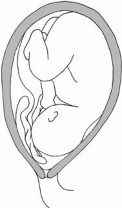
Fig. 19.1
Cord presentation.CORD PRESENTATION AND CORD PROLAPSE
441

Fig. 19.2
Cord prolapse.
CHAPTER 19
Emergencies442
Management
- Cord blood results are influenced by mode of delivery, gestational age, and analgesia during labour.
Other books
Expectation (Ghost Targets, #2) by Pogue, Aaron
Living With No Regrets by Jayton Young
Revenge of the Lawn Gnomes by R. L. Stine
Dawn of a New Age by Rick Bentsen
Sweet Spot by Lucy Felthouse
Guía de la Biblia. Nuevo Testamento by Isaac Asimov
The Fatal Fashione by Karen Harper
The Wedding by Buchanan, Lexi
Comeback by Richard Stark