i bc27f85be50b71b1 (55 page)
Read i bc27f85be50b71b1 Online
Authors: Unknown


178
AClITE CARE HANDBOOK FOR PHYSICAL THERAPISTS
Figure 3-5. Garden classification of sllbcapitai fractures. I, 2 nondispJaced;
=
3, 4 displaced. (With permission from A Unwin, K Jones /edsJ. Emergency
=
Orthopaedics and Trauma. Boston: Butterworth-Hei"emann, 1995;193.)
oral diaphysis and can occur with intertrochanteric or shaft fractures.18 lnter- and subtrochanteric fractures are summarized In Chapter Appendix Table 3-AA and are shown in Figure 3-6.
Femoral Shaft Fracture
Femoral shaft fractures result from high-energy trauma and are
often associated with major abdominal or pelvic injuries. Femoral
shaft fractures can be accompanied by life-threatening systemic
complications, such as hypovolemia, shock, or fat emboli. Often,
there is significant bleeding into the thigh with hematoma formation. Hip dislocation and patellar fracture frequently accompany femoral shaft fractures" Chapter Appendix Table 3-A.S lists femoral shaft fracture management.
Clinical Tip
• The uninvolved leg or a single crutch can be used to
assist the involved lower leg in and out of bed .
• The patient should avoid rotation of and pivoting on
the lower extremity after intramedullary rod placement, as
microrotation of the intramedullary rod can occur and
place stress on the fixation device.
Distal Femoral Fractures
Fractures of the distal femur are supracondylar (involving the distal
femur shaft only), Imicolldylar (involving one femoral condyle), ;'Iter-


MUSCUtOSKELHAL SYSTEM
179
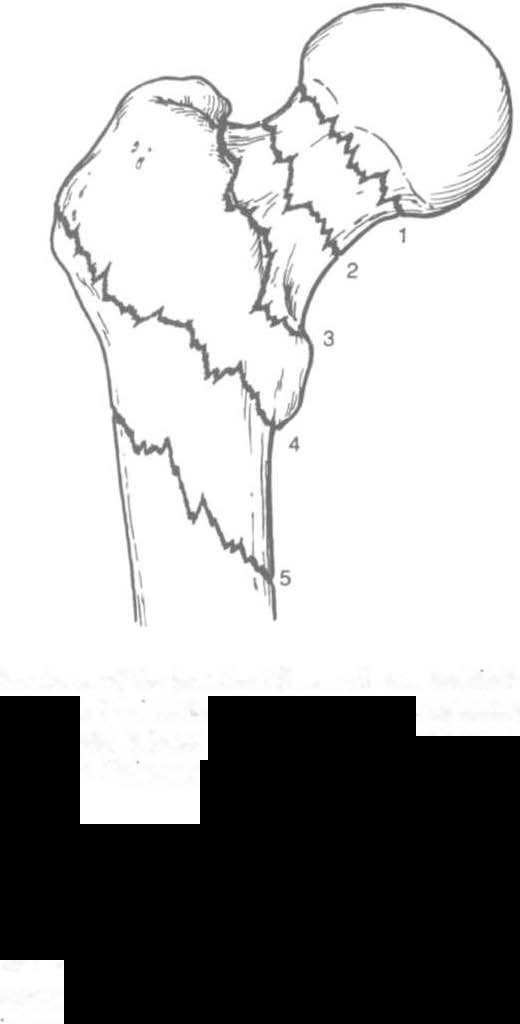
Figure 3-6. fmc/ures o( tbe /JroxUlIl1I (emur. (I = subcapltai; 2 = transcervicdi; 3 = b,1S()Cervica/; 4 = m/ertrocballleric; .5 = subtrochanteric.) (With permissi(J1l (rom A U",£11I1, K JOlles /cds/. emergency Orthopaedics and Trauma.
Bostoll: ButterllJortb-llememmm, 1995;190.)
cOlldy/ar (involving both condyles), or a combination of these.
Involvement of the articular surface of the knee joim complicates the
fracture. Typically, this type of fracture is caused by high-energy
trauma to the femur or a direct force that drives the tibia cranially
into the mtercondylar fossa. Distal femur fractures are often accompanied by mjury to localized skin, muscle, and joint ligaments and cartilage with great potential for decreased knee joint mechanics.2o
The management of distal femur fractures is summarized in Chapter
Appendix Table 3-A.6. Complications of femur fracture include
malunion, nonunion, infection, knee pain, and decreased knee ROM.21
Patellar Fractllre
A patellar fracture results from direct trauma to the patella from a fall,
blow, or MVA (e.g., dashboard injury). The knee is often flexed during
injury, as the patella fractures against the femur. Patellar fracture may
also occur from sudden forceful quadriceps comracrion, as in sports or


