Ross & Wilson Anatomy and Physiology in Health and Illness (199 page)
Read Ross & Wilson Anatomy and Physiology in Health and Illness Online
Authors: Anne Waugh,Allison Grant
Tags: #Medical, #Nursing, #General, #Anatomy

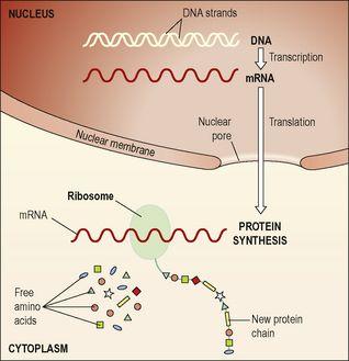
Figure 17.5
The relationship between DNA, RNA and protein synthesis.
Messenger ribonucleic acid (mRNA)
mRNA is a single-stranded chain of nucleotides synthesised in the nucleus from the appropriate gene, whenever the cell requires to make the protein for which that gene codes. There are three main differences between the structures of RNA and DNA:
•
it is single instead of double stranded
•
it contains ribose as its sugar instead of deoxyribose
•
it uses uracil instead of thymine as a base.
Using the DNA as a template, a piece of mRNA is made from the gene to be used. This process is called
transcription
. The mRNA then leaves the nucleus through the nuclear pores and carries its information to the ribosomes in the cytoplasm.
Transcription
Because the code is buried within the DNA molecule, the first step is to open up the helix to expose the bases. Only the gene to be transcribed is opened; the remainder of the chromosome remains coiled. Opening up the helix exposes both base strands, but the enzyme that makes the mRNA uses only one of them, so the mRNA molecule is single, not double stranded. As the enzyme moves along the opened DNA strand, reading its code, it adds the complementary base to the mRNA. Therefore, if the DNA base is cytosine, guanine is added to the mRNA molecule (and vice versa); if it is thymine, adenine is added; if it is adenine, uracil is added (remember there is no thymine in RNA, but uracil instead) (
Fig. 17.6
). When the enzyme gets to a ‘stop’ signal, it terminates synthesis of the mRNA molecule, and the mRNA is released. The DNA is zipped up again by other enzymes, and the mRNA then leaves the nucleus.
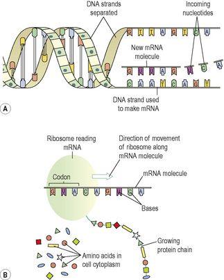
Figure 17.6
Transcription (A) and translation (B).
Translation
Translation is synthesis of the final protein using the information carried on mRNA. It takes place on ribosomes (
p. 29
) in the cytoplasm and on rough endoplasmic reticulum. First, the mRNA attaches to the ribosome. The ribosome then ‘reads’ the base sequence of the mRNA (
Fig. 17.6
).
Because proteins are built from up to 20 different amino acids, it is not possible to use the four bases individually in a simple one-to-one code. To give enough options, the base code in RNA is read in triplets, giving a possible 64 base combinations, which allows a coded instruction for each amino acid as well as other codes, e.g. stop and start instructions. Each of these specific triplet sequences is called a
codon
; for example, the base sequence ACA (adenine, cytosine, adenine) codes for the amino acid cysteine.
The first
codon
is a start codon, which initiates protein synthesis. The ribosome slides along the mRNA, reading the codons and adding the appropriate amino acids to the growing protein molecule as it goes. The ribosome continues assembling the new protein molecule until it arrives at a
stop codon
, at which point it terminates synthesis and releases the new protein. Some new proteins are used within the cell itself, and others are exported, e.g. insulin synthesised by pancreatic β-islet cells is released into the bloodstream.
Gene expression
Although all cells (except gametes and red blood cells) have an identical set of genes, each cell type uses only those genes related directly to its own particular function. For example, the only cell type
containing
haemoglobin is the red blood cell, although all body cells carry the haemoglobin gene. This selective gene expression is controlled by various regulatory substances, and the genes not needed by the cell are kept switched off.
Cell division
Learning outcomes
After studying this section, you should be able to:
explain the mechanism of DNA replication
compare and contrast the processes of mitosis and meiosis
describe the basis of genetic diversity from generation to generation.
Most body cells are capable of division, even in adulthood. Cell division usually leads to production of two identical diploid daughter cells,
mitosis
(
p. 31
) and is important in body growth and repair. Production of gametes is different in that the daughter cells have only half the normal chromosome number – 23 instead of 46, i.e. they are haploid. Gametes are produced by a form of cell division called
meiosis
. DNA replication takes place before mitosis and meiosis.
DNA replication
DNA is the only biological molecule capable of self-replication. Mistakes in copying may lead to production of non-functioning or poorly functional cells, or cells that do not respond to normal cell controls (this could lead to the development of a tumour). Accurate copying of DNA is therefore essential.
The initial step in DNA replication is the unfolding of the double helix and the unzipping of the two strands to expose the bases, as happens in transcription. Both strands of the parent DNA molecule are copied. The enzyme responsible for DNA replication moves along the base sequence on each strand, reading the genetic code and adding the complementary base to the newly forming chain. This means that each strand of opened bases becomes a double strand and the end result is two identical DNA molecules (
Fig. 17.7
). As each new double strand is formed, other enzymes cause it to twist and coil back into its normal highly folded form.


