Pediatric Examination and Board Review (33 page)
Read Pediatric Examination and Board Review Online
Authors: Robert Daum,Jason Canel

FIGURE 17-1.
See color plates.
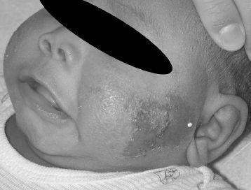
FIGURE 17-2.
See color plates.
4.
A 6-month-old male boy presents for evaluation of an erythematous plaque on the cheek, with a few small vesicles, moist surface, and yellow crust. The patient’s mother reports that this rash just developed in the last day and she has noticed the child scratching at it. Which of the following diagnostic tests is positive (
Figure 17-2
)?
(A) Tzanck smear
(B) Gram stain
(C) potassium hydroxide preparation
(D) Wright’s stain
(E) direct fluorescent antibody (DFA) assay
5.
The best management is
(A) mupirocin ointment to the rash 3 times a day
(B) acyclovir ointment 5-6 times a day
(C) IV acyclovir for 21 days
(D) clotrimazole cream twice daily
(E) griseofulvin suspension for 10 days
6.
The next child you see is a 6-month-old presenting for routine well-child care. You notice a few comedones on the child’s cheeks and upper back (
Figure 17-3
). The best management is
(A) tretinoin gel daily
(B) benzoyl peroxide gel daily
(C) testosterone and dehydroepiandrosterone sulfate (DHEAS) levels
(D) curettage
(E) hydrocortisone cream daily
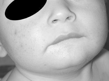
FIGURE 17-3.
See color plates.
7.
One week later this patient’s 3-year-old cousin presents with widespread erythema and scaling around the mouth, neck, and upper chest, revealing red moist skin under the scale. Rare fragile blisters are noted. He is clearly uncomfortable and hasn’t been drinking well. His lips are dry and he is febrile (
Figure 17-4
). He has not been on medication. The most likely diagnosis is
(A) toxic epidermal necrolysis
(B) Stevens-Johnson syndrome
(C) staphylococcal scalded skin syndrome
(D) toxic shock syndrome
(E) erythema multiforme
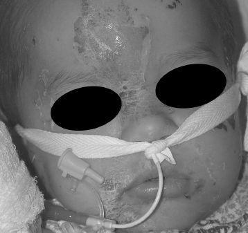
FIGURE 17-4.
See color plates.
8.
What is the best diagnostic test?
(A) culture of the blister fluid
(B) Gram stain of the blister fluid
(C) blood culture
(D) frozen section of sloughing skin
(E) viral culture from the base of the blister
9.
Staphylococcal scalded skin syndrome is caused by
(A) an allergic reaction to bacteria
(B) superantigens
(C) an exfoliatoxin
(D) a pyrogenic toxin
(E) reaction to antibiotics
10.
Full-thickness skin necrosis, severe mucosal involvement, and skin pain is characteristic of
(A) scarlet fever
(B) toxic epidermal necrolysis
(C) staphylococcal scalded skin syndrome
(D) bullous impetigo
(E) erythema multiforme
ANSWERS
1.
(E)
The timing of this rash, the patient’s otherwise good health, and the description of the lesions are all suggestive of transient neonatal pustular melanosis. This benign neonatal eruption classically presents in the first week of life, with lesions in multiple stages including vesicles, pustules, collarettes of scale, and hyperpigmented macules. It is more common in dark-skinned infants. Smear of the contents of a pustule demonstrates neutrophils and can be diagnostic. Erythema toxicum neonatorum is another common benign neonatal eruption, but it tends to spare the palms and soles and demonstrates blotchy erythema with central minute pustules. A smear of the contents of the pustule demonstrates eosinophils. Multinucleated giant cells are seen on Tzanck smears of herpetic lesions. An infant with neonatal herpes infection may appear sick, with lethargy and poor feeding. An infant with a presentation limited to skin may be asymptomatic, however, and develop neurologic symptoms and signs later. This diagnosis, then, is an important exclusion. The lesions are often eroded. Congenital candidiasis can present as pustules but typically presents in the first 24 hours of life and is more widespread with confluentappearing lesions and scaling. Scabies can cause an intense rash in infants, but the description of pustules leaving hyperpigmented macules and rims of scale is less classic for scabies in which vesicles, crusts, and dermatitic changes may be more notable.
2.
(C)
Transient neonatal pustular melanosis is a benign neonatal eruption for which no treatment is necessary. New lesions cease to develop during the first week of life. The postinflammatory hyperpigmentation may linger for weeks to months. Recognition of this common neonatal rash is important to spare these infants from unnecessary procedures, such as a septic workup and unnecessary drugs. Cultures of these pustules, both bacterial and viral, are sterile.
3.
(D)
One month of age is a typical time to present with neonatal acne. This is a common finding in both boys and girls. The lesions typically affect the face and are characterized by erythematous papules and pustules without comedones. This is a selflimited eruption and no treatment is indicated. The dark patches that are noted refer to leftover hyperpigmentation from the transient neonatal pustulosis.
4.
(B)
A Gram stain will demonstrate gram-positive cocci, confirming the diagnosis of impetigo. Impetigo is a common skin infection in children, caused by both
Staphylococcus aureus
and group A streptococci. The presence of yellow crust and the location near an orifice are suggestive of this diagnosis. More rarely, herpes simplex can present in infants and is important to recognize and rule out. The typical lesions are grouped vesicles on a red base, without the characteristic yellow crust of impetigo. A Tzanck smear or DFA can be used to raise clinical suspicion about a herpetic lesion. Tinea corporis might also be considered. These lesions are classically more annular with scale and not crust.
5.
(A)
Topical treatment of impetigo with an antibacterial, such as mupirocin, is often adequate when the lesions are localized. Treatment of herpes simplex virus (HSV) in patients of this age is somewhat controversial. Infants younger than 3 months of age are treated aggressively with IV antivirals and are evaluated for central nervous system (CNS) manifestations of herpes infection. There may still be concern at 6 months of age that a local infection can disseminate or that the eruption represents reactivation of a congenital infection.
6.
(B)
Acneiform lesions at 6 months of age represent infantile acne, which is more common in boys. They are characterized by comedones, and likely are androgen driven. However, testosterone and DHEAS levels are typically normal and do not necessarily need to be measured unless the child demonstrates other signs of virilization. Treatment with benzoyl peroxide is often helpful. Tretinoin is generally reserved for the most recalcitrant cases. Curettage is not indicated. Hydrocortisone may make the condition worse.
7.
(C)
Periorificial and flexural involvement with fragile blisters, peeling skin, and moist denuded exposed skin is typical of staphylococcal scalded skin syndrome, caused by an exfoliatoxinproducing strain of
S aureus
phage group 2. Toxic epidermal necrolysis (TEN) and Stevens-Johnson syndrome are most commonly drug-induced hypersensitivity reactions and mucosal involvement is prominent. Toxic shock syndrome does not cause blistering. Erythema multiforme is a hypersensitivity reaction sometimes associated with HSV and presents with targetoid lesions.
8.
(D)
The gold standard for diagnosis of staphylococcal scalded skin syndrome is demonstration of blister formation within the stratum granulosum of the epidermis, in contrast to TEN in which epidermis is necrotic in full thickness. Cultures from the blisters and denuded skin are sterile. A blood culture is only rarely positive. The highest yield of the inciting bacteria is from the nasopharynx or conjunctiva.
9.
(C)
Staphylococcal scalded skin syndrome is caused by
S aureus
strains producing exfoliatoxin; these strains usually belong to phage group 2. Pyrogenic toxins are implicated in scarlet fever caused by group A streptococci.
10.
(B)
Full-thickness skin necrosis, severe mucosal involvement, and skin pain is typical of TEN. This is usually a drug-induced hypersensitivity reaction, which is distinguished from Stevens-Johnson syndrome and erythema multiforme by the extent of surface area involvement. Ophthalmologic involvement is also common and must be sought.
 S
S
UGGESTED
R
EADING