The Washington Manual Internship Survival Guide (32 page)
Read The Washington Manual Internship Survival Guide Online
Authors: Thomas M. de Fer,Eric Knoche,Gina Larossa,Heather Sateia
Tags: #Medical, #Internal Medicine

Pneumothorax
•
Pneumothorax is usually an urgent consult;
it is an emergent consult if the patient is receiving positive pressure ventilation or if there is suspicion of a tension pneumothorax
.
•
Common symptoms include dyspnea and chest pain. Careful examination for signs of tension pneumothorax (deviation of the trachea to the opposite side, muffled or absent breath sounds on the affected side, and hypotension) must be performed. If there are no signs of tension pneumothorax, an upright chest X-ray should be obtained.
•
Pertinent information:
• What is the patient’s exam and how does he/she look?
• Which side is affected?
• Is there a CXR?
• Is the patient on positive pressure ventilation?
• Is this a tension pneumothorax?
•
Treatment: Options vary as to the size and the physiologic impact of the pneumothorax:
•
If a tension pneumothorax is found and the patient is decompensating, immediately place a large-bore (14G or 16G) angiocatheter in the second interspace above the rib at the midclavicular line of the affected side
. A chest tube tray and a 28 French chest tube should be brought to the bedside so that the consultant may place the tube as soon as they arrive.
• If the patient is on positive pressure ventilation, the patient should be monitored closely until tube thoracostomy is performed. If the patient decompensates, immediately place a large (14G or 16G) angiocatheter in the second interspace at the midclavicular line of the affected side.
• Expectant management with serial chest radiographs may suffice in a young, asymptomatic patient with a small pneumothorax who is not on positive pressure ventilation.
• Most patients may require open tube thoracostomy or percutaneous tube thoracostomy (a 16 French Thal-Quick tube).
Pleural Effusion
•
Pleural effusions are generally an elective or semi-urgent consult, unless the patient is short of breath, in which case an emergent consult is warranted.
•
Common symptoms include dyspnea and chest pain. Effusions can result from a wide spectrum of benign, malignant, and inflammatory conditions.
•
Pertinent information:
• Is the patient short of breath and/or having trouble breathing? Depending on the size of the effusion and the patient’s pulmonary status, patients will differ in their symptomatology.
• Are there any CXR, chest CT, or US studies that document the effusion?
• Is it getting bigger?
• Has a thoracentesis been done? If so, what did the Gram stain/culture, pH, glucose, amylase, lactate dehydrogenase, protein levels, cell count, and cytology show of the fluid?
•
Treatment: Options vary depending on the etiology and the character of the pleural effusion.
• Pleural effusions generally need to be drained via thoracentesis, open tube thoracostomy, or placement of an implantable
long-term catheter that allows repeated drainage of recurrent pleural effusions.
• Fluid should be sent off for Gram stain and culture, cytology, cell count, and biochemical analyses (pH, glucose, amylase, LDH, protein) to help discriminate between an exudative and a transudative effusion.
• Additional options include streptokinase for loculated effusions or pleurodesis for recurrent effusions.
Perirectal Abscess
•
Generally this is an
urgent consult unless the patient is septic, then it is an emergency!
Remember that there is no such thing as an unimportant abscess. It should always be evaluated by a surgeon.
•
Pertinent information: Is the patient diabetic or immunosuppressed? If so, then they are far more likely to die or to have greater morbidity from this disease. Is the patient febrile, and do they have a leukocytosis?
•
Physical exam:
• Where is it (relative to the scrotum/vagina and anus)?
• How far does the erythema and induration extend? If the stigmata of infection extend out from the anus, then the patient may have
Fournier gangrene
, which is a surgical emergency!
• Has a CT scan been obtained?
•
Treatment options:
•
Incision and drainage
either at the bedside or in the operating room.
•
IV antibiotics
.
• Fournier’s gangrene will necessitate wide debridement in the operating room with massive irrigation of the affected areas.
PRESSURE ULCERS
•
Generally these are elective or urgent consults unless the patient is septic.
•
Pressure ulcers result from prolonged pressure to soft tissue over bony prominences. Most commonly they occur in immobile patients over the occiput, sacrum, greater trochanter, and heels.
•
Pertinent information:
• Is the patient septic?
• Is the patient diabetic or immunosuppressed? Has the area been irradiated in the past? What is the patient’s nutritional status?
• Is the patient immobile? What is the extent and etiology of the patient’s immobility?
• What is the duration of the ulcer?
• What is their current wound care management?
•
Physical exam: How does it look? Where is it? How deep is it? Is there any erythema, induration, or fluctuance around it? Do you see any exposed bone?
•
Treatment options:
• Most superficial pressure ulcers heal spontaneously when the pressure is relieved; however, this can be a lengthy process requiring over 6 months.
• Local wound care and optimization of nutrition are key for ulcer healing. Urinary and fecal continence needs to be maintained to prevent maceration and skin breakdown.
• Simple closure, split-thickness skin grafting, or musculocutaneous flaps are possible, but often not successful unless the pressure can be removed.
ABCs of Critical Care
22
KEYS TO INTENSIVE CARE UNIT SURVIVAL
•
Transfer notes with key details of the patient’s past medical history and course are always helpful. The primary physician, receiving physician, and the patient’s family members should be notified. Note any details or special situations that need attention and/or follow-up. This should always be in addition to a conversation between physicians.
•
Admitting a patient to an ICU can be intimidating, but keep in mind the ABCs (Airway, Breathing, Circulation) and focus on stabilizing the patient.
•
Be nice to the nurses, respiratory therapists, and all other ancillary staff during your stay. They can often make very useful suggestions, catch things you miss, and be immensely helpful to you in critical situations. They can be the difference between an enjoyable and miserable experience!
•
Ask (and keep asking) if you have questions or problems
. Mistakes from inexperience in critically ill patients can have catastrophic consequences.
•
Always treat the patient, not the numbers.
•
It’s always a good idea to make rounds on patients and follow up on labs several times a day even if things seem stable.
•
Daily ICU notes should include ventilator settings, I/Os, pulmonary artery catheter measurements, medications and drips (e.g., antibiotics, sedatives, vasopressors, inotropes), nutritional status, and documentation of all hardware (e.g., central venous catheters, arterial lines, endotracheal tube, feeding tubes).
•
References to have nearby at all times:
• Lederman RJ.
Tarascon Internal Medicine and Critical Care Pocketbook
. 5th ed. Burlington, MA: Jones & Bartlett Learning; 2011.
• Kollef MH, Isakow W.
The Washington Manual of Critical Care
. Philadelphia, PA: Lippincott Williams & Wilkins; 2011.
• Kollef MH, Micek ST. Critical care. In: Foster C, Mistry NF, Peddi PF, Sharma S, eds.
Washington Manual of Medical Therapeutics
. 33rd ed. Philadelphia, PA: Lippincott Williams & Wilkins; 2010. 34th edition out in 2013.
VENTILATORS
Suggestions for Initial Ventilator Settings
Basic Settings
•
Mode:
• AC/VC: Volume control. Minimal minute ventilation is set, often best mode to start with. Tidal volume is set. Airway pressures are dependent on compliance.
• SIMV + PSV (i.e., synchronous intermittent mandatory ventilation with pressure support ventilation): Similar to AC except patient-triggered breaths are delivered with pressure support instead of full tidal volume.
• PCV: Pressure-controlled ventilation. Airway pressures are set and the tidal volume delivered depends on compliance.
•
Tidal volume: 6 mL/kg IBW for lung protective ventilation in ARDS patients; otherwise start with 6 to 9 mL/kg.
•
Rate: 10 to 15 breaths/min.
•
FIO
2
: 1.00, then titrate down (goal to get FIO
2
to ≤0.60 as quickly as possible).
•
PEEP: 5 cm H
2
O (monitor for auto-PEEP, especially with obstructive lung disease).
Advanced Settings
•
Ask for help before you change these.
•
Inspiratory flow: 50 to 60 L/min. With COPD may need >100 L/min.
•
I:E ratio: 1:2–1:3. This must be set with PCV.
•
Peak and plateau pressures: Goal plateau pressure <32 cm H
2
O; prefer peak pressure <45 cm H
2
O.
Ventilator Adjustments
•
PO
2
of 60 mm Hg or greater is generally sufficient. Oxygenation is most affected by mean air pressure. Adjustments to FIO
2
and PEEP can help increase oxygenation. Remember that the
oxygen saturations and PO
2
can be discordant
, need to verify that they are consistent.

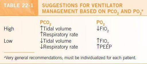
•
PCO
2
is regulated by minute ventilation. Increasing the respiratory rate or tidal volume increases minute ventilation and decreases PCO
2.
•
See
Table 22-1
for suggested adjustments.
Worsening Oxygenation
The knee-jerk impulse is to turn up the FIO
2
.
Don’t panic!
Approach the problem in a stepwise manner:
•
Is there a ventilator problem?
•
Is the ET tube in the correct position or has it migrated? Check the positioning (e.g., 25 cm at the lip) of the ET tube and look at the most recent chest radiograph.
•
Is there a cuff leak or kink in the ET tube? The respiratory therapist can help evaluate.
•
Recheck the ventilator settings. Have there been inadvertent changes?
•
Is there an obstruction in the ET tube (e.g., mucous plug)? Pass the suction catheter to evaluate.
•
Is it a patient-related problem (e.g., biting, agitation)? Sedation may be needed.
•
Always consider pneumothorax. Listen on both sides and consider a CXR.
•
Is the underlying problem worsening? Is the patient fluid overloaded or is there bronchospasm? Is PE a possibility? Has ventilator-associated pneumonia developed?
•
Is the patient oversedated? Do you need to rethink the mode of ventilation? Check the PEEP at end expiration. Hypoxemia can result from a loss of PEEP.
Weaning Parameters
