Examination Medicine: A Guide to Physician Training (83 page)
Read Examination Medicine: A Guide to Physician Training Online
Authors: Nicholas J. Talley,Simon O’connor
Tags: #Medical, #Internal Medicine, #Diagnosis

Table 16.23
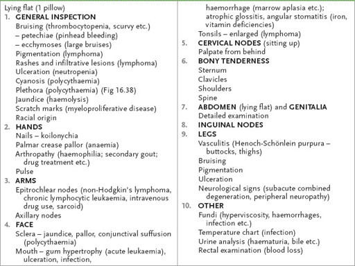
Haematological system examination
1.
If necessary, position the patient as for a gastrointestinal examination. Make sure he is fully undressed. Look for bruising, pigmentation, cyanosis, jaundice, scratch marks (owing to myeloproliferative disease or lymphoma) and leg ulceration (see below). Also note the presence of frontal bossing and the racial origin of the patient (thalassaemia is more common in people of Asian or Greek background).
2.
Pick up the patient’s hands. Look at the nails for koilonychia (spoon-shaped nails, which are rarely seen today and which indicate iron deficiency) and the changes of vasculitis. Pale palmar creases indicate anaemia (usually the haemoglobin level is <90 g/L). Evidence of arthropathy may be important – for example, rheumatoid arthritis and Felty’s syndrome, recurrent haemarthroses in bleeding disorders or secondary gout in myeloproliferative disorders.
3.
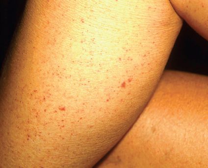
Examine the epitrochlear nodes. Do this by placing your palm under the patient’s elbow – your thumb will be placed over the appropriate area (proximal and slightly anterior to the medial epicondyle). A palpable node is usually pathological and may indicate non-Hodgkin’s lymphoma. Note any arm bruising. Remember petechiae (
Fig 16.37
) are pinhead haemorrhages, whereas ecchymoses are larger bruises. Palpable purpura suggests vasculitis, dysglobulinaemia or bacteraemia.
FIGURE 16.37
Petechiae. J G Marks, J J Miller.
Lookingbill and Marks’ principles of dermatology
, 4th edn. Fig 17.1. Saunders, Elsevier, 2006, with permission.
4.
Go to the axillae and palpate the axillary nodes. Do this by raising the patient’s arm and placing your fingers as high as possible in the axilla. Then position the patient’s forearm comfortably over your own forearm. Use your left hand for the patient’s right axilla, and vice versa. There are four main areas: central, lateral (above and lateral), pectoral (most medial) and subscapular (most inferior).
5.
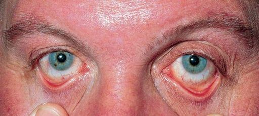
Look at the patient’s face. Inspecting the eyes, note jaundice, pallor or haemorrhage of the sclera, or the injected sclera of polycythaemia.
HINT
Anaemia is most accurately detected by pulling down one of the patient’s lower eyelids and comparing the colour of the anterior part of the conjunctiva with the pearly white of the palpebral conjunctiva more posteriorly. The anterior conjunctiva should be distinctly more red in colour unless significant anaemia (<100 g/L) is present.
6.
Examine the mouth. Note gum hypertrophy (differential diagnosis includes acute myeloid leukaemia – especially monocytic – and scurvy), ulceration, infection (e.g.
Candida
), haemorrhage, atrophic glossitis (secondary to iron, vitamin B
12
or folate deficiency) and angular stomatitis. Look for tonsillar and adenoid enlargement (Waldeyer’s ring).
7.
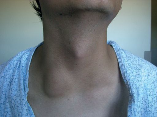
Now sit the patient up. Examine the cervical nodes from behind (
Fig 16.39
). There are seven groups: submental, submandibular, jugular chain, posterior triangle, postauricular, preauricular and occipital. Then feel the supraclavicular area from the front. Tap the spine with your fist for bony tenderness (which can be caused by an enlarging marrow – e.g. in myeloma, carcinoma). Also gently press the sternum, clavicles and shoulders for bony tenderness.
FIGURE 16.39
Supraclavicular lymphadenopathy.
8.
Lay the patient flat again. Examine the abdomen, particularly for splenomegaly (see
Table 16.21
) and hepatomegaly (see
Tables 16.17
and
16.22
).
9.
Spring the hips for pelvic tenderness.
10.
Palpate the inguinal nodes. There are two groups – along the inguinal ligament and along the femoral vessels. Remember to feel the testes and ask to do a rectal examination (e.g. for melaena).
11.
Examine the legs. Note particularly leg ulcers, which may occur with hereditary spherocytosis, sickle cell syndromes, thalassaemia, macroglobulinaemia and Felty’s syndrome. Ask to examine the legs from a neurological aspect for evidence of vitamin B
12
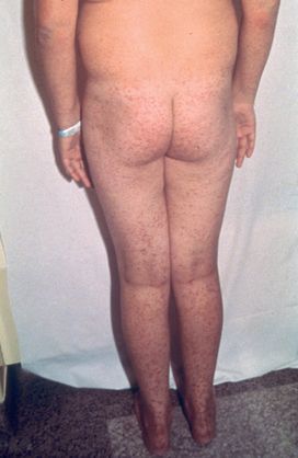
deficiency. Remember, hypothyroidism and lead poisoning can cause anaemia and peripheral neuropathy. Do not miss Henoch-Schönlein purpura over the buttocks and legs (
Fig 16.41
).
FIGURE 16.41
Henock-Schönlein purpura. N Talley, S O’Connor,
Clinical examination
, 7th edn. Fig 21.15. Elsevier, 2013, with permission. From F S McDonald (ed.)
Mayo Clinic images in internal medicine
, with permission. Copyright Mayo Clinic Scientific Press and CRC Press.
12.
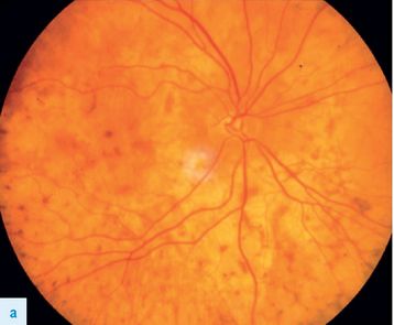
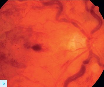
Finally, ask to examine the fundi (engorged retinal vessels, papilloedema, haemorrhages etc.) (
Fig 16.40a and b
) and look at the temperature chart. Causes of generalised lymphadenopathy are presented in
Table 16.24
.
Table 16.24
Causes of generalised lymphadenopathy
FIGURE 16.38
Plethora. A V Hoffbrand, J E Pettite.
Color atlas of clinical hematology
, 3rd edn. p. 248. Mosby, Elsevier, 2000, with permission.
1.
Lymphoma (rubbery and firm)
2.
Leukaemia (chronic lymphocytic leukaemia, acute lymphoblastic leukaemia particularly)
3.
Malignant disease (metastases or reactive changes usually causing asymmetrical, very firm nodes)
4.
Infections – viral (e.g. cytomegalovirus, HIV, infectious mononucleosis (glandular fever)), bacterial (e.g. tuberculosis, brucellosis), protozoal (e.g. toxoplasmosis)
5.
Connective tissue diseases – rheumatoid arthritis, SLE
6.
Infiltrations – sarcoidosis
7.
Drugs – phenytoin (pseudolymphoma)
FIGURE 16.40
Fundus changes in haematological disorders (a) ‘Leopard skin’ appearance due to choroidal infiltration in chronic leukaemia.
(b) Retinal and gross venous dilatation and segmentation in hyperviscocity. J J Kanski, Retinal vascular disease.
Clinical opthalmology: a systematic approach
. Elsevier, 2011, with permission.
The endocrine system
The thyroid gland
‘This 40-year-old woman has noticed some discomfort in her neck. Please examine her.’
Method (see
Table 16.25
)
