Read Your Inner Fish: A Journey Into the 3.5-Billion-Year History of the Human Body Online
Authors: Neil Shubin
Your Inner Fish: A Journey Into the 3.5-Billion-Year History of the Human Body (14 page)


If we follow the gill arches from an embryo to an adult, we can trace the origins of jaws, ears, larynx, and throat. Bones, muscles, nerves, and arteries all develop inside these gill arches.
This fundamental blueprint of heads helps us make sense of one of the apocryphal tales in anatomy. In 1820, so the story goes, Johannes Goethe was walking through the Jewish cemetery in Vienna when he spotted the decomposing skeleton of a ram. The vertebrae were exposed and above them lay a damaged skull. Goethe, in a moment of epiphany, saw that the breaks in the skull made it look like a gnarled mess of vertebrae. To Goethe, this revealed the essential pattern within: the head is made up of vertebrae that fused and grew a vault to hold our brains and sense organs. This was a revolutionary idea because it linked heads and bodies as two versions of the same fundamental plan. The notion must have been in the drinking water in the early 1800s because other people, among them Lorenz Oken, allegedly came up with virtually the same idea in a similar setting.
Goethe and Oken were both picking up something very profound, although they could not have known it at the time. Our body is segmented, and this pattern is most clearly seen in our vertebrae. Each vertebra is a block that represents a segment of our body. The organization of our nerves is also segmental, correlating closely with the pattern of the vertebrae. Nerves exit the spinal cord to supply the body. The segmental configuration is obvious when you look at the levels of the spinal cord that are associated with each part of our body. For example, the muscles in our legs are supplied by nerves that exit from lower parts of the spinal cord than those that supply our arms. Heads may not look it, but they also contain a very deep segmental pattern. Our arches define segments of bones, muscles, arteries, and nerves. Look in the adult, and you won’t see this pattern. We see it only in the embryo.
Our skulls lose all overt evidence of their segmental origins as we go from embryo to adult. The plate-like bones of our skulls form over our gill arches, and the muscles, nerves, and arteries, which all had a very simple segmental pattern early on, are rewired to make our adult heads.
Knowing something about development can help us predict where to look for what is missing in children who have certain birth defects. For example, children born with first arch syndrome have a tiny jaw and nonfunctioning ears with no malleus or incus bone. Missing are structures that normally would have formed from the first arch.
The arches are the road map for major chunks of the skull, from the most complicated cranial nerves to the muscles, arteries, bones, and glands inside. The arches are also a guide to something else: our very deep connection with sharks.
OUR INNER SHARK

The take-home message of many a lawyer joke is that lawyers are an especially voracious kind of shark. Teaching embryology during one of the recurring vogues for these jokes, I remember thinking that the joke is on all of us. We’re all modified sharks—or, worse, there is a lawyer inside each of us.
As we’ve seen, much of the secret of heads lies in the arches, the swellings that gave us the road map for the complicated cranial nerves and key structures inside the head. Those insignificant-looking swellings and indentations have captured the imagination of anatomists for 150 years, because they look like the gill slits in the throat regions of fish and sharks.
Fish embryos have these bulges and indentations, too. In fish, the indentations ultimately open up to form the spaces between the gills where water flows. In us, the indentations normally seal over. In abnormal cases, gill slits fail to close and remain open as pouches or cysts. A branchial cyst, for example, is often a benign fluid-filled cyst that forms in an open pouch inside the neck; the pouch is created by the failure of the third or fourth arch to close. Rarely, children are born with an actual vestige of an ancient gill arch cartilage, a little rod that represents a gill bar from the third arch. In these instances, my surgical colleagues are operating on an inner fish that unfortunately has come back to bite us.
Every head on every animal from a shark to a human shares those four arches in development. The richness of the story lies in what happens inside each arch. Here, we can make a point-by-point comparison between our heads and those of sharks.

The gill region of a developing human and a developing shark look the same early on.
Look at the first arch in a human and a shark, and you find a very similar state of affairs: jaws. The major difference is that humans’ first arch also forms some ear bones, which we do not see in sharks. Unsurprisingly, the cranial nerve that supplies the jaws of humans and sharks is the first arch nerve, the trigeminal nerve.
The cells inside the second gill arch divide, change, and give rise to a bar of cartilage and muscle. In us, the bar of cartilage breaks up to form one of the three bones of our middle ear (the stapes) and some other small structures at the base of the head and throat. One of these bones, called the hyoid, assists us in swallowing. Take a gulp, listen to music, and thank the structures that form from your second arch.
In a shark, the second arch rod breaks up to form two bones that support the jaws: a lower one that compares with our hyoid and an upper one that supports the upper jaw. If you have ever watched a great white shark try to chomp something—a diver in a cage, for example—you have probably noticed that the upper jaw can extend and retract as the shark bites. The upper bone of this second arch is part of the lever system of bones that rotate to make that possible. That upper bone is remarkable in another way, too. It compares with one of the bones in our middle ear: the stapes. Bones that support the upper and lower jaws in sharks are used in us to swallow and hear.
As for the third and fourth arches, we find that many of the structures we use to talk and swallow are, in sharks, parts of tissues that support the gills. The muscles and cranial nerves we use to swallow and talk move the gills in sharks and fish.
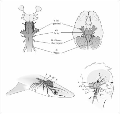
At first glance, our cranial nerves (bottom right) appear different from those of a shark (bottom left). But look closely and you will find profound similarities. Virtually all of our nerves are present in sharks. The parallels go deeper still: equivalent nerves in sharks and humans supply similar structures, and they even exit the brain in the same order (top left and right).
Our head may look incredibly complicated, but it is built from a simple and elegant blueprint. There is a pattern common to every skull on earth, whether it belongs to a shark, a bony fish, a salamander, or a human. The discovery of this pattern was a major accomplishment of nineteenth-century anatomy, a time when anatomists were putting embryos of all kinds of species under the microscope. In 1872, the Oxford anatomist Francis Maitland Balfour first saw the basic plan of heads when he looked at sharks and saw the bulges, the gill arches, and the structures inside. Unfortunately, he died soon after in a mountaineering accident in the Swiss Alps. He was only in his thirties.
GILL ARCH GENES
During the first three weeks after conception, whole batteries of genes are turned on and off in our gill arches and throughout the tissues that will become our future brain. These genes instruct cells to make the different portions of our head. Think of each region of our head as gaining a genetic address that makes it distinctive. Modify this genetic address and we can modify the kinds of structures that develop there.
For example, a gene known as
Otx
is active in the front region, where the first gill arch forms. Behind it, toward the back of the head, a number of so-called
Hox
genes are active. Each gill arch has a different complement of
Hox
genes active in it. With this information, we can make a map of our gill arches and the constellation of genes active in making each.
Now we can do experiments: change the genetic address of one gill arch into that of another. Take a frog embryo, turn off some genes, make the genetic signals similar in the first and second arches, and you end up with a frog that has two jaws: a mandible develops where a hyoid bone would normally be. This shows the power of the genetic addresses in making our gill arches. Change the address, and you change the structures in the arch. The power of this approach is that we can now experiment with the basic design of heads: we can manipulate the identity of the gill arches almost at will, by changing the activity of the genes inside.
TRACING HEADS: FROM HEADLESS WONDERS TO OUR HEADED ANCESTORS